Want to create or adapt books like this? Learn more about how Pressbooks supports open publishing practices.

8.4 Nerve Impulses
Created by CK-12 Foundation/Adapted by Christine Miller
When Lightning Strikes
This amazing cloud-to-surface lightning occurred when a difference in electrical charge built up in a cloud relative to the ground. When the buildup of charge was great enough, a sudden discharge of electricity occurred. A nerve impulse is similar to a lightning strike. Both a nerve impulse and a lightning strike occur because of differences in electrical charge, and both result in an electric current.
Generating Nerve Impulses
A nerve impulse , like a lightning strike, is an electrical phenomenon. A nerve impulse occurs because of a difference in electrical charge across the plasma membrane of a neuron. How does this difference in electrical charge come about? The answer involves ions , which are electrically-charged atoms or molecules .
Resting Potential
When a neuron is not actively transmitting a nerve impulse, it is in a resting state, ready to transmit a nerve impulse. During the resting state, the sodium-potassium pump maintains a difference in charge across the cell membrane of the neuron. The sodium-potassium pump is a mechanism of active transport that moves sodium ions (Na+) out of cells and potassium ions (K+) into cells. The sodium-potassium pump moves both ions from areas of lower to higher concentration, using energy in ATP and carrier proteins in the cell membrane. The video below, “Sodium Potassium Pump” by Amoeba Sisters, describes in greater detail how the sodium-potassium pump works. Sodium is the principal ion in the fluid outside of cells, and potassium is the principal ion in the fluid inside of cells. These differences in concentration create an electrical gradient across the cell membrane, called resting potential . Tightly controlling membrane resting potential is critical for the transmission of nerve impulses.
Sodium Potassium Pump, Amoeba Sisters, 2020.
Action Potential
A nerve impulse is a sudden reversal of the electrical gradient across the plasma membrane of a resting neuron. The reversal of charge is called an action potential . It begins when the neuron receives a chemical signal from another cell or some other type of stimulus . If the stimulus is strong enough to reach threshold , an action potential will take place is a cascade along the axon.
This reversal of charges ripples down the axon of the neuron very rapidly as an electric current, which is illustrated in the diagram below (Figure 8.4.2). A nerve impulse is an all-or-nothing response depending on if the stimulus input was strong enough to reach threshold. If a neuron responds at all, it responds completely. A greater stimulation does not produce a stronger impulse.
In neurons with a myelin sheath on their axon, ions flow across the membrane only at the nodes between sections of myelin. As a result, the action potential appears to jump along the axon membrane from node to node, rather than spreading smoothly along the entire membrane. This increases the speed at which the action potential travels.
Transmitting Nerve Impulses
The place where an axon terminal meets another cell is called a synapse . This is where the transmission of a nerve impulse to another cell occurs. The cell that sends the nerve impulse is called the presynaptic cell , and the cell that receives the nerve impulse is called the postsynaptic cell .
Some synapses are purely electrical and make direct electrical connections between neurons. Most synapses, however, are chemical synapses. Transmission of nerve impulses across chemical synapses is more complex.
Chemical Synapses
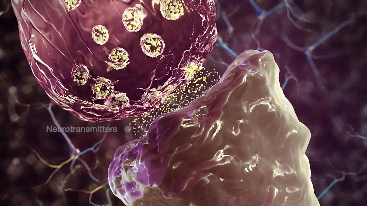
At a chemical synapse, both the presynaptic and postsynaptic areas of the cells are full of molecular machinery that is involved in the transmission of nerve impulses. As shown in Figure 8.4.3, the presynaptic area contains many tiny spherical vessels called synaptic vesicles that are packed with chemicals called neurotransmitters . When an action potential reaches the axon terminal of the presynaptic cell, it opens channels that allow calcium to enter the terminal. Calcium causes synaptic vesicles to fuse with the membrane, releasing their contents into the narrow space between the presynaptic and postsynaptic membranes. This area is called the synaptic cleft . The neurotransmitter molecules travel across the synaptic cleft and bind to receptors , which are proteins embedded in the membrane of the postsynaptic cell.
Neurotransmitters and Receptors
There are more than a hundred known neurotransmitters, and more than one type of neurotransmitter may be released at a given synapse by a presynaptic cell. For example, it is common for a faster-acting neurotransmitter to be released, along with a slower-acting neurotransmitter. Many neurotransmitters also have multiple types of receptors to which they can bind. Receptors, in turn, can be divided into two general groups: chemically gated ion channels and second messenger systems.
- When a chemically gated ion channel is activated, it forms a passage that allows specific types of ions to flow across the cell membrane. Depending on the type of ion, the effect on the target cell may be excitatory or inhibitory .
- When a second messenger system is activated, it starts a cascade of molecular interactions inside the target cell. This may ultimately produce a wide variety of complex effects, such as increasing or decreasing the sensitivity of the cell to stimuli, or even altering gene transcription.
The effect of a neurotransmitter on a postsynaptic cell depends mainly on the type of receptors that it activates, making it possible for a particular neurotransmitter to have different effects on various target cells. A neurotransmitter might excite one set of target cells, inhibit others, and have complex modulatory effects on still others, depending on the type of receptors. However, some neurotransmitters have relatively consistent effects on other cells. Consider the two most widely used neurotransmitters, glutamate and GABA (gamma-aminobutyric acid). Glutamate receptors are either excitatory or modulatory in their effects, whereas GABA receptors are all inhibitory in their effects in adults.
Problems with neurotransmitters or their receptors can cause neurological disorders. The disease myasthenia gravis , for example, is caused by antibodies from the immune system blocking receptors for the neurotransmitter acetylcholine in postsynaptic muscle cells. This inhibits the effects of acetylcholine on muscle contractions, producing symptoms, such as muscle weakness and excessive fatigue during simple activities. Some mental illnesses (including depression ) are caused, at least in part, by imbalances of certain neurotransmitters in the brain. One of the neurotransmitters involved in depression is thought to be serotonin , which normally helps regulate mood, among many other functions. Some antidepressant drugs are thought to help alleviate depression in many patients by normalizing the activity of serotonin in the brain.
8.4 Summary
- A nerve impulse is an electrical phenomenon that occurs because of a difference in electrical charge across the plasma membrane of a neuron.
- The sodium-potassium pump maintains an electrical gradient across the plasma membrane of a neuron when it is not actively transmitting a nerve impulse. This gradient is called the resting potential of the neuron.
- An action potential is a sudden reversal of the electrical gradient across the plasma membrane of a resting neuron. It begins when the neuron receives a chemical signal from another cell or some other type of stimulus. The action potential travels rapidly down the neuron’s axon as an electric current and occurs in three stages: Depolarization, Repolarization and Recovery.
- A nerve impulse is transmitted to another cell at either an electrical or a chemical synapse . At a chemical synapse, neurotransmitter chemicals are released from the presynaptic cell into the synaptic cleft between cells. The chemicals travel across the cleft to the postsynaptic cell and bind to receptors embedded in its membrane.
- There are many different types of neurotransmitters. Their effects on the postsynaptic cell generally depend on the type of receptor they bind to. The effects may be excitatory, inhibitory, or modulatory in more complex ways. Both physical and mental disorders may occur if there are problems with neurotransmitters or their receptors.
8.4 Review Questions
- Define nerve impulse.
- What is the resting potential of a neuron, and how is it maintained?
- Explain how and why an action potential occurs.
- Outline how a signal is transmitted from a presynaptic cell to a postsynaptic cell at a chemical synapse.
- What generally determines the effects of a neurotransmitter on a postsynaptic cell?
- Identify three general types of effects that neurotransmitters may have on postsynaptic cells.
- Explain how an electrical signal in a presynaptic neuron causes the transmission of a chemical signal at the synapse.
- The flow of which type of ion into a neuron results in an action potential? How do these ions get into the cell? What does this flow of ions do to the relative charge inside the neuron compared to the outside?
- Name three neurotransmitters.
8.4 Explore More
Action Potentials, Teacher’s Pet, 2018.
TED Ed| What is depression? – Helen M. Farrell, Parta Learning, 2017.
5 Weird Involuntary Behaviors Explained!, It’s Okay To Be Smart, 2015.
Attributions
Figure 8.4.1
Lightening/ Purple Lightning, Dee Why by Jeremy Bishop on Unsplash is used under the Unsplash License (https://unsplash.com/license).
Figure 8.4.2
Action Potential by CNX OpenStax, Biology on Wikimedia Commons is used under a CC BY 4.0 (https://creativecommons.org/licenses/by/4.0/deed.en) license.
Figure 8.4.3
Chemical_synapse_schema_cropped by Looie496 created file (adapted from original from US National Institutes of Health, National Institute on Aging) is in the public domain (https://en.wikipedia.org/wiki/Public_domain).
Amoeba Sisters. (2020, January 29). Sodium potassium pump. YouTube. https://www.youtube.com/watch?v=7NY6XdPBhxo&feature=youtu.be
CNX OpenStax. (2016, May 27) Figure 4 The action potential is conducted down the axon as the axon membrane depolarizes, then repolarizes [digital image]. In Open Stax, Biology (Section 35.2). OpenStax CNX. https://cnx.org/contents/[email protected]:cs_Pb-GW@6/How-Neurons-Communicate
It’s Okay To Be Smart. (2015, January 26). 5 Weird involuntary behaviors explained! YouTube. https://www.youtube.com/watch?v=ZE8sRMZ5BCA&feature=youtu.be
Mayo Clinic Staff. (n.d.). Depression (major depressive disorder) [online article]. MayoClinic.org. https://www.mayoclinic.org/diseases-conditions/depression/symptoms-causes/syc-20356007
Mayo Clinic Staff. (n.d.). Myasthenia gravis [online article]. MayoClinic.org. https://www.mayoclinic.org/diseases-conditions/myasthenia-gravis/symptoms-causes/syc-20352036
National Institute on Aging. (2006, April 8). Alzheimers disease: Unraveling the mystery. National Institutes of Health. https://www.nia.nih.gov/ ( archived version )
Parta Learning. (2017, December 8). TED Ed| What is depression? – Helen M. Farrell. YouTube. https://www.youtube.com/watch?v=rBcU_apy0h8&t=291s
Teacher’s Pet. (2018, August 26). Action potentials. YouTube. https://www.youtube.com/watch?v=FEHNIELPb0s&feature=youtu.be
A signal transmitted along a nerve fiber.
An atom or molecule with a net electric charge due to the loss or gain of one or more electrons.
The smallest particle of an element that still has the properties of that element.
A molecule is an electrically neutral group of two or more atoms held together by chemical bonds.
A functional unit of the nervous system that transmits nerve impulses; also called a nerve cell.
A solute pump that pumps potassium into cells while pumping sodium out of cells, both against their concentration gradients. This pumping is active and occurs at the ratio of 2 potassium for every 3 calcium.
The semipermeable membrane surrounding the cytoplasm of a cell.
The movement of ions or molecules across a cell membrane into a region of higher concentration, assisted by enzymes and requiring energy.
A complex organic chemical that provides energy to drive many processes in living cells, e.g. muscle contraction, nerve impulse propagation, and chemical synthesis. Found in all forms of life, ATP is often referred to as the "molecular unit of currency" of intracellular energy transfer.
The difference in electrical charge across the plasma membrane of a neuron that is not actively transmitting a nerve impulse.
Reversal of electrical charge across the membrane of a resting neuron that travels down the axon of the neuron as a nerve impulse.
Something that triggers a behavior or other response.
The critical level to which a membrane potential must be depolarized to initiate an action potential.
The place where the axon terminal of a neuron transmits a chemical or electrical signal to another cell.
The cell that sends the nerve impulse.
The cell that receives the nerve impulse.
These membrane-bound organelles store various neurotransmitters that are released at the synapse. The release is regulated by a voltage-dependent calcium channel. Vesicles are essential for propagating nerve impulses between neurons and are constantly recreated by the cell.
A type of chemical that transmits signals from the axon of a neuron to another cell across a synapse.
A space that separates two neurons. It forms a junction between two or more neurons and helps nerve impulse pass from one neuron to the other.
A protein on a cell membrane or inside of a cell that binds with a hormone, neurotransmitter, or other chemical signal to produce a response.
A neurotransmitter that will have excitatory effects on the neuron, meaning it will increase the likelihood that a neuron will fire an action potential.
A neurotransmitter that decreases the likelihood that a neuron will fire an action potential.
A chemical that nerve cells use to send signals to other cells. It is by a wide margin the most abundant excitatory neurotransmitter in the vertebrate nervous system.
A naturally occurring amino acid that works as a neurotransmitter in your brain. Neurotransmitters function as chemical messengers. GABA is considered an inhibitory neurotransmitter because it blocks, or inhibits, certain brain signals and decreases activity in your nervous system.
An antibody, also known as an immunoglobulin, is a large, Y-shaped protein produced mainly by plasma cells that is used by the immune system to neutralize pathogens such as pathogenic bacteria and viruses.
An organic chemical that functions in the brain and body of many types of animals (and humans) as a neurotransmitter—a chemical message released by nerve cells to send signals to other cells, such as neurons, muscle cells and gland cells.
A neurotransmitter. It has a popular image as a contributor to feelings of well-being and happiness, though its actual biological function is complex and multifaceted, modulating cognition, reward, learning, memory, and numerous physiological processes such as vomiting and vasoconstriction.
Human Biology Copyright © 2020 by Christine Miller is licensed under a Creative Commons Attribution-NonCommercial 4.0 International License , except where otherwise noted.
Share This Book

- school Campus Bookshelves
- menu_book Bookshelves
- perm_media Learning Objects
- login Login
- how_to_reg Request Instructor Account
- hub Instructor Commons
- Download Page (PDF)
- Download Full Book (PDF)
- Periodic Table
- Physics Constants
- Scientific Calculator
- Reference & Cite
- Tools expand_more
- Readability
selected template will load here
This action is not available.
13.5: Nerve Impulse
- Last updated
- Save as PDF
- Page ID 13276

How does a nervous system signal move from one cell to the next?
It literally jumps by way of a chemical transmitter. Notice the two cells are not connected, but separated by a small gap. The synapse. The space between a neuron and the next cell.
Nerve Impulses
Nerve impulses are electrical in nature. They result from a difference in electrical charge across the plasma membrane of a neuron. How does this difference in electrical charge come about? The answer involves ions , which are electrically charged atoms or molecules.
Resting Potential
When a neuron is not actively transmitting a nerve impulse, it is in a resting state, ready to transmit a nerve impulse. During the resting state, the sodium-potassium pump maintains a difference in charge across the cell membrane (see Figure below). It uses energy in ATP to pump positive sodium ions (Na + ) out of the cell and potassium ions (K + ) into the cell. As a result, the inside of the neuron is negatively charged compared to the extracellular fluid surrounding the neuron. This is due to many more positively charged ions outside the cell compared to inside the cell. This difference in electrical charge is called the resting potential.

Action Potential
A nerve impulse is a sudden reversal of the electrical charge across the membrane of a resting neuron. The reversal of charge is called an action potential. It begins when the neuron receives a chemical signal from another cell. The signal causes gates in sodium ion channels to open, allowing positive sodium ions to flow back into the cell. As a result, the inside of the cell becomes positively charged compared to the outside of the cell. This reversal of charge ripples down the axon very rapidly as an electric current (see Figure below).

In neurons with myelin sheaths, ions flow across the membrane only at the nodes between sections of myelin. As a result, the action potential jumps along the axon membrane from node to node, rather than spreading smoothly along the entire membrane. This increases the speed at which it travels.
The place where an axon terminal meets another cell is called a synapse . The axon terminal and other cell are separated by a narrow space known as a synaptic cleft (see Figure below). When an action potential reaches the axon terminal, the axon terminal releases molecules of a chemical called a neurotransmitter . The neurotransmitter molecules travel across the synaptic cleft and bind to receptors on the membrane of the other cell. If the other cell is a neuron, this starts an action potential in the other cell.

- A nerve impulse begins when a neuron receives a chemical stimulus.
- The nerve impulse travels down the axon membrane as an electrical action potential to the axon terminal.
- The axon terminal releases neurotransmitters that carry the nerve impulse to the next cell.
- Define resting potential and action potential.
- Explain how resting potential is maintained
- Describe how an action potential occurs.
- What is a synapse?
If you're seeing this message, it means we're having trouble loading external resources on our website.
If you're behind a web filter, please make sure that the domains *.kastatic.org and *.kasandbox.org are unblocked.
To log in and use all the features of Khan Academy, please enable JavaScript in your browser.
Biology library
Course: biology library > unit 33.
- Anatomy of a neuron
- Overview of neuron structure and function
- The membrane potential
- Electrotonic and action potentials
- Saltatory conduction in neurons
- Neuronal synapses (chemical)
The synapse
- Neurotransmitters and receptors
- Q & A: Neuron depolarization, hyperpolarization, and action potentials
- Overview of the functions of the cerebral cortex
- Neurons communicate with one another at junctions called synapses . At a synapse, one neuron sends a message to a target neuron—another cell.
- Most synapses are chemical ; these synapses communicate using chemical messengers. Other synapses are electrical ; in these synapses, ions flow directly between cells.
- At a chemical synapse, an action potential triggers the presynaptic neuron to release neurotransmitters . These molecules bind to receptors on the postsynaptic cell and make it more or less likely to fire an action potential.
Introduction
Electrical or chemical transmission.
- Some people thought that signaling across a synapse involved the flow of ions directly from one neuron into another—electrical transmission.
- Other people thought it depended on the release of a chemical from one neuron, causing a response in the receiving neuron—chemical transmission.
Overview of transmission at chemical synapses
Excitatory and inhibitory postsynaptic potentials.
- In some cases, the change makes the target cell more likely to fire its own action potential. In this case, the shift in membrane potential is called an excitatory postsynaptic potential , or EPSP .
- In other cases, the change makes the target cell less likely to fire an action potential and is called an inhibitory post-synaptic potential , or IPSP .
Spatial and temporal summation
- The integration of postsynaptic potentials that occur in different locations—but at about the same time—is known as spatial summation .
- The integration of postsynaptic potentials that occur in the same place—but at slightly different times—is called temporal summation .
Signal termination
Chemical synapses are flexible, electrical synapses, works cited.
- David E. Sadava, David M. Hillis, H. Craig Heller, and May Berenbaum, "How Do Neurons Communicate with Other Cells?" In Life: The Science of Biology , 9th ed. (Sunderland: Sinauer Associates, 2009), 961.
- Alberto E. Pereda, "Electrical Synapses and Their Functional Interactions with Chemical Synapses," Nature Reviews Neuroscience 15 (2014): 250-263, http://dx.doi.org/10.1038/nrn3708 .
Suggestions for further reading
Want to join the conversation.
- Upvote Button navigates to signup page
- Downvote Button navigates to signup page
- Flag Button navigates to signup page


- school Campus Bookshelves
- menu_book Bookshelves
- perm_media Learning Objects
- login Login
- how_to_reg Request Instructor Account
- hub Instructor Commons
- Download Page (PDF)
- Download Full Book (PDF)
- Periodic Table
- Physics Constants
- Scientific Calculator
- Reference & Cite
- Tools expand_more
- Readability
selected template will load here
This action is not available.
11.4: Neuronal Communication
- Last updated
- Save as PDF
- Page ID 22338

- Whitney Menefee, Julie Jenks, Chiara Mazzasette, & Kim-Leiloni Nguyen
- Reedley College, Butte College, Pasadena City College, & Mt. San Antonio College via ASCCC Open Educational Resources Initiative
By the end of this section, you will be able to:
- Explain the events that occur during the conduction of nerve impulses
- Differentiate the nerve impulse propagation in saltatory and continuous conduction
- Describe the components of synapses and compare electrical and chemical synapses
Having looked at the components of nervous tissue, and the basic anatomy of the nervous system, next comes an understanding of how nervous tissue is capable of communicating within the nervous system. Neurons communicate with other neurons, muscles or glands through the generation and conduction of nerve impulses. These nerve impulses represent changes in the electrical properties of the neuronal cell membrane. All cells have an electrical charge associated with their membrane. However, neurons and other cells are able to change their electrical charge by moving ions across the membrane. In this section, you will look at the basics of neuronal communication, mainly focusing on the conduction of the nerve impulse.
Conduction of Nerve Impulses
Neurons possess electrical excitability, which is the ability to respond to a stimulus by generating a nerve impulse. In the majority of cases, the dendrites of a neuron are the place where local changes in the membrane electrical properties happen through synapses. Dendrites receive stimuli from the external environment (e.g. somatic sensory neurons) or internal environment (e.g. visceral sensory neurons, motor neurons or interneurons). The amount of change in the membrane electrical charge is determined by the strength of the stimulus that causes it. For example, a needle pricking a fingertip will result in a bigger stimulus compared to a blunt object touching the same fingertip. Once a stimulus (or multiple stimuli) produces a significant change in the membrane electrical properties of a dendrite and reaches a predetermined threshold , then a nerve impulse (also called action potential) occurs. An action potential is generated at the axon hillock of a neuron and progresses rapidly along the axon's plasma membrane to reach the targets (a second neuron, a muscle or a gland). This movement of an action potential along the axon is called propagation . While the stimuli can be weak or strong, the action potential follows a All-or-None Law by which it always has the same strength (referred to as amplitude) independently of the stimulus. This minimizes the possibility that information will be lost along the way. The only way to modulate the response is through the frequency of action potentials - how many action potentials reach the target in a set amount of time. A bigger stimulus will produce a series (or train) of action potentials that are close together, while a weak stimulus will produce sparse action potentials.
The speed of an action potential is influenced by the diameter of the axon and by its myelination. The larger the diameter of the axon, the faster the action potential will be conducted. Myelinated axons are able to carry action potentials faster than unmyelinated axons. As discussed in the previous section, myelinating cells (oligodendrocytes in the CNS and Schwann cells in the PNS) wrap around axons forming the myelin. Nodes of Ranvier are gaps between segments of myelin. The electrical charges of the action potential can "jump" from one gap to another one, thus allowing a faster speed of the action potential. This progression of a nerve impulse is called saltatory conduction . However, in unmyelinated axons, one side of the axon is not covered by myelin and the electrical charges move along the entire axonal membrane, thus taking longer to reach their target. This progression of a nerve impulse is called continuous conduction . Once the action potential reaches the axon terminal, it is either transported as electrical charge into the next cell or transformed into a chemical signal, depending on the type of synapse that the synaptic end bulb is forming with its target.
Neurons and their targets form synapses. The neuron that generates and conducts the action potential to the target is called a presynaptic cell . The target cell receiving the action potential is called a postsynaptic cell . While the presynaptic cell is always a neuron (because only neurons have axons and can form a synapse), the postsynaptic cell can be a neuron or another type of cell such as skeletal, cardiac or smooth muscle cells, or glands. In Figure \(\PageIndex{1}\), a presynaptic neuron forms synapses with two postsynaptic neurons. The nerve impulse (or signal) travels from a presynaptic neuron to a postsynaptic cell. If the postsynaptic cell is a neuron, a new action potential might be generated in the postsynaptic neuron and reach its postsynaptic targets.

There are two types of connections between electrically active cells: electrical synapses and chemical synapses. In an electrical synapse , there is a direct connection between the presynaptic and postsynaptic cells and the connection is formed by gap junctions. Thus, the electrical charges of an action potential can pass directly from one cell to the next. If one cell delivers an action potential in an electrical synapse, the joined cell will also generate an action potential because the electrical charges will pass between the cells (Figure \(\PageIndex{2}\)). Although representing the minority of synapses, electrical synapses are found throughout the nervous system. These synapses also occur between excitable cells other than neurons, for example between smooth muscle cells in the intestines and cardiac muscle cells in the heart.

Chemical synapses involve the transmission of chemical information from one cell to the next and they represent the majority of the synapses found within the nervous system. In a chemical synapse , a chemical signal called a neurotransmitter , is released from the presynaptic cell and it affects the postsynaptic cell. There are many different types of neurotransmitters, for example acetylcholine, serotonin, dopamine, adrenaline, glutamate, etc. Each neurotransmitter has its own specific receptor on the postsynaptic membrane. Chemical synapses can then be classified based on the neurotransmitter that the cells use to communicate (for example glutamatergic synapses use glutamate). Different neurotransmitters and different receptors will determine the overall response to the stimulus. All chemical synapses have common characteristics, which can be summarized in this list and are shown in Figure \(\PageIndex{3}\):
- synaptic end bulb of presynaptic neuron
- neurotransmitter (packaged in vesicles)
- synaptic cleft
- receptors for neurotransmitter
- postsynaptic membrane of postsynaptic neuron
The synaptic end bulbs of chemical synapses are filled with vesicles containing one type of neurotransmitter. When an action potential reaches the axon terminals, the vesicles merge with the cell membrane at the synaptic end bulb, releasing the neurotransmitter through exocytosis into the small gap between the cells, known as the synaptic cleft . Once in the synaptic cleft, the neurotransmitter diffuses the short distance to the postsynaptic membrane and can interact with neurotransmitter receptors. Receptors are specific for the neurotransmitter, and the two fit together like a key and lock. One neurotransmitter binds to its receptor and will not bind to receptors for other neurotransmitters, making the binding a specific chemical event. The binding of the neurotransmitter to its receptor causes a brief electrical change across the postsynaptic membrane. The change depends on the type of neurotransmitter receptor. Changes in the postsynaptic cell membrane can cause a nerve impulse to begin in the postsynaptic cell or inhibit the generation of an action potential. The flow of information is unidirectional: from the presynaptic cell to the postsynaptic cell. After its release in a chemical synapse, neurotransmitters need to be removed from the synaptic cleft to ensure the propagation of new synaptic signals.

DISORDERS OF THE...
Nervous System: Alzheimer's and Parkinson's Disease
The underlying cause of some neurodegenerative diseases, such as Alzheimer’s and Parkinson’s, appears to be related to proteins—specifically, to proteins behaving badly. One of the strongest theories of what causes Alzheimer’s disease is based on the accumulation of beta-amyloid plaques, dense conglomerations of a protein that is not functioning correctly. Parkinson’s disease is linked to an increase in a protein known as alpha-synuclein that is toxic to the cells of the substantia nigra nucleus in the midbrain.
For proteins to function correctly, they are dependent on their three-dimensional shape. The linear sequence of amino acids folds into a three-dimensional shape that is based on the interactions between and among those amino acids. When the folding is disturbed, and proteins take on a different shape, they stop functioning correctly. But the disease is not necessarily the result of functional loss of these proteins; rather, these altered proteins start to accumulate and may become toxic. For example, in Alzheimer’s, the hallmark of the disease is the accumulation of these amyloid plaques in the cerebral cortex. The term coined to describe this sort of disease is “proteopathy” and it includes other diseases. Creutzfeld-Jacob disease, the human variant of the prion disease known as mad cow disease in the bovine, also involves the accumulation of amyloid plaques, similar to Alzheimer’s. Diseases of other organ systems can fall into this group as well, such as cystic fibrosis or type 2 diabetes. Recognizing the relationship between these diseases has suggested new therapeutic possibilities. Interfering with the accumulation of the proteins, and possibly as early as their original production within the cell, may unlock new ways to alleviate these devastating diseases.
CAREER CONNECTIONS
Neurophysiologist
Understanding how the nervous system works could be a driving force in your career. Studying neurophysiology is a very rewarding path to follow. It means that there is a lot of work to do, but the rewards are worth the effort.
The career path of a research scientist can be straightforward: college, graduate school, postdoctoral research, academic research position at a university. A Bachelor’s degree in science will get you started, and for neurophysiology that might be in biology, psychology, computer science, engineering, or neuroscience. But the real specialization comes in graduate school. There are many different programs out there to study the nervous system, not just neuroscience itself. Most graduate programs are doctoral, meaning that a Master’s degree is not part of the work. These are usually considered five-year programs, with the first two years dedicated to course work and finding a research mentor, and the last three years dedicated to finding a research topic and pursuing that with a near single-mindedness. The research will usually result in a few publications in scientific journals, which will make up the bulk of a doctoral dissertation. After graduating with a Ph.D., researchers will go on to find specialized work called a postdoctoral fellowship within established labs. In this position, a researcher starts to establish their own research career with the hopes of finding an academic position at a research university.
Other options are available if you are interested in how the nervous system works. Especially for neurophysiology, a medical degree might be more suitable so you can learn about the clinical applications of neurophysiology and possibly work with human subjects. An academic career is not a necessity. Biotechnology firms are eager to find motivated scientists ready to tackle the tough questions about how the nervous system works so that therapeutic chemicals can be tested on some of the most challenging disorders such as Alzheimer’s disease or Parkinson’s disease, or spinal cord injury.
Others with a medical degree and a specialization in neuroscience go on to work directly with patients, diagnosing and treating mental disorders. You can do this as a psychiatrist, a neuropsychologist, a neuroscience nurse, or a neurodiagnostic technician, among other possible career paths.
Concept Review
The basis of the electrical signal within a neuron is the action potential that propagates down the axon. For a neuron to generate an action potential, it needs to receive input from another source, either another neuron or a sensory stimulus. That input will result in a change in the electrical properties of the cell membrane, based on the strength of the stimulus. Once the stimulus is strong enough, it will generate an action potential that travels along the axon to the synaptic end bulb.
The diameter of the axon and the presence or absence of myelin determines how fast the action potential is conducted down the axon. A larger diameter and the presence of a myelin sheath will ensure a fast propagation of the action potential.
Synapses are the contacts between neurons, which can either be chemical or electrical in nature. Chemical synapses are far more common. At a chemical synapse, neurotransmitter is released from the presynaptic element and diffuses across the synaptic cleft. The neurotransmitter binds to a receptor protein and causes a change in the postsynaptic membrane (the PSP). The neurotransmitter must diffuse, be inactivated or removed from the synaptic cleft so that the stimulus is limited in time.
Review Questions
Q. At an electrical synapse, what do the presynaptic and postsynaptic cells communicate through?
A. neurotransmitters
B. neurotransmitter receptors
C. nodes of Ranvier
D. gap junctions
Q. Which of the following axons would propagate an action potential faster than the others?
A. myelinated, large diameter, axons
B. myelinated, small diameter, axons
C. unmyelinated, large diameter, axons
D. unmyelinated, small diameter, axons
Contributors and Attributions
OpenStax Anatomy & Physiology (CC BY 4.0). Access for free at https://openstax.org/books/anatomy-and-physiology

Want to create or adapt books like this? Learn more about how Pressbooks supports open publishing practices.
80 8.4 Nerve Impulses
Created by CK-12 Foundation/Adapted by Christine Miller

When Lightning Strikes
This amazing cloud-to-surface lightning occurred when a difference in electrical charge built up in a cloud relative to the ground. When the buildup of charge was great enough, a sudden discharge of electricity occurred. A nerve impulse is similar to a lightning strike. Both a nerve impulse and a lightning strike occur because of differences in electrical charge, and both result in an electric current.
Generating Nerve Impulses
A nerve impulse , like a lightning strike, is an electrical phenomenon. A nerve impulse occurs because of a difference in electrical charge across the plasma membrane of a neuron. How does this difference in electrical charge come about? The answer involves ions , which are electrically-charged atoms or molecules .
Resting Potential
When a neuron is not actively transmitting a nerve impulse, it is in a resting state, ready to transmit a nerve impulse. During the resting state, the sodium-potassium pump maintains a difference in charge across the cell membrane of the neuron. The sodium-potassium pump is a mechanism of active transport that moves sodium ions (Na+) out of cells and potassium ions (K+) into cells. The sodium-potassium pump moves both ions from areas of lower to higher concentration, using energy in ATP and carrier proteins in the cell membrane. The video below, “Sodium Potassium Pump” by Amoeba Sisters, describes in greater detail how the sodium-potassium pump works. Sodium is the principal ion in the fluid outside of cells, and potassium is the principal ion in the fluid inside of cells. These differences in concentration create an electrical gradient across the cell membrane, called resting potential . Tightly controlling membrane resting potential is critical for the transmission of nerve impulses.
Sodium Potassium Pump, Amoeba Sisters, 2020.
Action Potential
A nerve impulse is a sudden reversal of the electrical gradient across the plasma membrane of a resting neuron. The reversal of charge is called an action potential . It begins when the neuron receives a chemical signal from another cell or some other type of stimulus . If the stimulus is strong enough to reach threshold , an action potential will take place is a cascade along the axon.
This reversal of charges ripples down the axon of the neuron very rapidly as an electric current, which is illustrated in the diagram below (Figure 8.4.2). A nerve impulse is an all-or-nothing response depending on if the stimulus input was strong enough to reach threshold. If a neuron responds at all, it responds completely. A greater stimulation does not produce a stronger impulse.

In neurons with a myelin sheath on their axon, ions flow across the membrane only at the nodes between sections of myelin. As a result, the action potential appears to jump along the axon membrane from node to node, rather than spreading smoothly along the entire membrane. This increases the speed at which the action potential travels.
Transmitting Nerve Impulses
The place where an axon terminal meets another cell is called a synapse . This is where the transmission of a nerve impulse to another cell occurs. The cell that sends the nerve impulse is called the presynaptic cell , and the cell that receives the nerve impulse is called the postsynaptic cell .
Some synapses are purely electrical and make direct electrical connections between neurons. Most synapses, however, are chemical synapses. Transmission of nerve impulses across chemical synapses is more complex.
Chemical Synapses
At a chemical synapse, both the presynaptic and postsynaptic areas of the cells are full of molecular machinery that is involved in the transmission of nerve impulses. As shown in Figure 8.4.3, the presynaptic area contains many tiny spherical vessels called synaptic vesicles that are packed with chemicals called neurotransmitters . When an action potential reaches the axon terminal of the presynaptic cell, it opens channels that allow calcium to enter the terminal. Calcium causes synaptic vesicles to fuse with the membrane, releasing their contents into the narrow space between the presynaptic and postsynaptic membranes. This area is called the synaptic cleft . The neurotransmitter molecules travel across the synaptic cleft and bind to receptors , which are proteins embedded in the membrane of the postsynaptic cell.

Neurotransmitters and Receptors
There are more than a hundred known neurotransmitters, and more than one type of neurotransmitter may be released at a given synapse by a presynaptic cell. For example, it is common for a faster-acting neurotransmitter to be released, along with a slower-acting neurotransmitter. Many neurotransmitters also have multiple types of receptors to which they can bind. Receptors, in turn, can be divided into two general groups: chemically gated ion channels and second messenger systems.
- When a chemically gated ion channel is activated, it forms a passage that allows specific types of ions to flow across the cell membrane. Depending on the type of ion, the effect on the target cell may be excitatory or inhibitory .
- When a second messenger system is activated, it starts a cascade of molecular interactions inside the target cell. This may ultimately produce a wide variety of complex effects, such as increasing or decreasing the sensitivity of the cell to stimuli, or even altering gene transcription.
The effect of a neurotransmitter on a postsynaptic cell depends mainly on the type of receptors that it activates, making it possible for a particular neurotransmitter to have different effects on various target cells. A neurotransmitter might excite one set of target cells, inhibit others, and have complex modulatory effects on still others, depending on the type of receptors. However, some neurotransmitters have relatively consistent effects on other cells. Consider the two most widely used neurotransmitters, glutamate and GABA (gamma-aminobutyric acid). Glutamate receptors are either excitatory or modulatory in their effects, whereas GABA receptors are all inhibitory in their effects in adults.
Problems with neurotransmitters or their receptors can cause neurological disorders. The disease myasthenia gravis , for example, is caused by antibodies from the immune system blocking receptors for the neurotransmitter acetylcholine in postsynaptic muscle cells. This inhibits the effects of acetylcholine on muscle contractions, producing symptoms, such as muscle weakness and excessive fatigue during simple activities. Some mental illnesses (including depression ) are caused, at least in part, by imbalances of certain neurotransmitters in the brain. One of the neurotransmitters involved in depression is thought to be serotonin , which normally helps regulate mood, among many other functions. Some antidepressant drugs are thought to help alleviate depression in many patients by normalizing the activity of serotonin in the brain.
8.4 Summary
- A nerve impulse is an electrical phenomenon that occurs because of a difference in electrical charge across the plasma membrane of a neuron.
- The sodium-potassium pump maintains an electrical gradient across the plasma membrane of a neuron when it is not actively transmitting a nerve impulse. This gradient is called the resting potential of the neuron.
- An action potential is a sudden reversal of the electrical gradient across the plasma membrane of a resting neuron. It begins when the neuron receives a chemical signal from another cell or some other type of stimulus. The action potential travels rapidly down the neuron’s axon as an electric current and occurs in three stages: Depolarization, Repolarization and Recovery.
- A nerve impulse is transmitted to another cell at either an electrical or a chemical synapse . At a chemical synapse, neurotransmitter chemicals are released from the presynaptic cell into the synaptic cleft between cells. The chemicals travel across the cleft to the postsynaptic cell and bind to receptors embedded in its membrane.
- There are many different types of neurotransmitters. Their effects on the postsynaptic cell generally depend on the type of receptor they bind to. The effects may be excitatory, inhibitory, or modulatory in more complex ways. Both physical and mental disorders may occur if there are problems with neurotransmitters or their receptors.
8.4 Review Questions
- Define nerve impulse.
- What is the resting potential of a neuron, and how is it maintained?
- Explain how and why an action potential occurs.
- Outline how a signal is transmitted from a presynaptic cell to a postsynaptic cell at a chemical synapse.
- What generally determines the effects of a neurotransmitter on a postsynaptic cell?
- Identify three general types of effects that neurotransmitters may have on postsynaptic cells.
- Explain how an electrical signal in a presynaptic neuron causes the transmission of a chemical signal at the synapse.
- The flow of which type of ion into a neuron results in an action potential? How do these ions get into the cell? What does this flow of ions do to the relative charge inside the neuron compared to the outside?
- Name three neurotransmitters.
8.4 Explore More
Action Potentials, Teacher’s Pet, 2018.
TED Ed| What is depression? – Helen M. Farrell, Parta Learning, 2017.
5 Weird Involuntary Behaviors Explained!, It’s Okay To Be Smart, 2015.
Attributions
Figure 8.4.1
Lightening/ Purple Lightning, Dee Why by Jeremy Bishop on Unsplash is used under the Unsplash License (https://unsplash.com/license).
Figure 8.4.2
Action Potential by CNX OpenStax, Biology on Wikimedia Commons is used under a CC BY 4.0 (https://creativecommons.org/licenses/by/4.0/deed.en) license.
Figure 8.4.3
Chemical_synapse_schema_cropped by Looie496 created file (adapted from original from US National Institutes of Health, National Institute on Aging) is in the public domain (https://en.wikipedia.org/wiki/Public_domain).
Amoeba Sisters. (2020, January 29). Sodium potassium pump. YouTube. https://www.youtube.com/watch?v=7NY6XdPBhxo&feature=youtu.be
CNX OpenStax. (2016, May 27) Figure 4 The action potential is conducted down the axon as the axon membrane depolarizes, then repolarizes [digital image]. In Open Stax, Biology (Section 35.2). OpenStax CNX. https://cnx.org/contents/[email protected]:cs_Pb-GW@6/How-Neurons-Communicate
It’s Okay To Be Smart. (2015, January 26). 5 Weird involuntary behaviors explained! YouTube. https://www.youtube.com/watch?v=ZE8sRMZ5BCA&feature=youtu.be
Mayo Clinic Staff. (n.d.). Depression (major depressive disorder) [online article]. MayoClinic.org. https://www.mayoclinic.org/diseases-conditions/depression/symptoms-causes/syc-20356007
Mayo Clinic Staff. (n.d.). Myasthenia gravis [online article]. MayoClinic.org. https://www.mayoclinic.org/diseases-conditions/myasthenia-gravis/symptoms-causes/syc-20352036
National Institute on Aging. (2006, April 8). Alzheimers disease: Unraveling the mystery. National Institutes of Health. https://www.nia.nih.gov/ ( archived version )
Parta Learning. (2017, December 8). TED Ed| What is depression? – Helen M. Farrell. YouTube. https://www.youtube.com/watch?v=rBcU_apy0h8&t=291s
Teacher’s Pet. (2018, August 26). Action potentials. YouTube. https://www.youtube.com/watch?v=FEHNIELPb0s&feature=youtu.be
A signal transmitted along a nerve fiber.
An atom or molecule with a net electric charge due to the loss or gain of one or more electrons.
The smallest particle of an element that still has the properties of that element.
A molecule is an electrically neutral group of two or more atoms held together by chemical bonds.
A functional unit of the nervous system that transmits nerve impulses; also called a nerve cell.
A solute pump that pumps potassium into cells while pumping sodium out of cells, both against their concentration gradients. This pumping is active and occurs at the ratio of 2 potassium for every 3 calcium.
The semipermeable membrane surrounding the cytoplasm of a cell.
The movement of ions or molecules across a cell membrane into a region of higher concentration, assisted by enzymes and requiring energy.
A complex organic chemical that provides energy to drive many processes in living cells, e.g. muscle contraction, nerve impulse propagation, and chemical synthesis. Found in all forms of life, ATP is often referred to as the "molecular unit of currency" of intracellular energy transfer.
The difference in electrical charge across the plasma membrane of a neuron that is not actively transmitting a nerve impulse.
Reversal of electrical charge across the membrane of a resting neuron that travels down the axon of the neuron as a nerve impulse.
Something that triggers a behavior or other response.
The critical level to which a membrane potential must be depolarized to initiate an action potential.
The place where the axon terminal of a neuron transmits a chemical or electrical signal to another cell.
The cell that sends the nerve impulse.
The cell that receives the nerve impulse.
These membrane-bound organelles store various neurotransmitters that are released at the synapse. The release is regulated by a voltage-dependent calcium channel. Vesicles are essential for propagating nerve impulses between neurons and are constantly recreated by the cell.
A type of chemical that transmits signals from the axon of a neuron to another cell across a synapse.
A space that separates two neurons. It forms a junction between two or more neurons and helps nerve impulse pass from one neuron to the other.
A protein on a cell membrane or inside of a cell that binds with a hormone, neurotransmitter, or other chemical signal to produce a response.
A neurotransmitter that will have excitatory effects on the neuron, meaning it will increase the likelihood that a neuron will fire an action potential.
A neurotransmitter that decreases the likelihood that a neuron will fire an action potential.
A chemical that nerve cells use to send signals to other cells. It is by a wide margin the most abundant excitatory neurotransmitter in the vertebrate nervous system.
A naturally occurring amino acid that works as a neurotransmitter in your brain. Neurotransmitters function as chemical messengers. GABA is considered an inhibitory neurotransmitter because it blocks, or inhibits, certain brain signals and decreases activity in your nervous system.
An antibody, also known as an immunoglobulin, is a large, Y-shaped protein produced mainly by plasma cells that is used by the immune system to neutralize pathogens such as pathogenic bacteria and viruses.
An organic chemical that functions in the brain and body of many types of animals (and humans) as a neurotransmitter—a chemical message released by nerve cells to send signals to other cells, such as neurons, muscle cells and gland cells.
A neurotransmitter. It has a popular image as a contributor to feelings of well-being and happiness, though its actual biological function is complex and multifaceted, modulating cognition, reward, learning, memory, and numerous physiological processes such as vomiting and vasoconstriction.
Human Biology Copyright © 2020 by Christine Miller is licensed under a Creative Commons Attribution-NonCommercial 4.0 International License , except where otherwise noted.
Share This Book

Nerve Impulses: the Key to Understanding the Brain
Impulses are the basis of mind..
Posted October 17, 2019
One of the greatest, relatively underappreciated, discoveries in all of science was the discovery of the nerve impulse in the 1930s by the British Lord Adrian. Adrian did win a Nobel Prize for his discovery in 1932, but scholars underestimated its implications, which go beyond the fact that four later Nobel Prizes were awarded for work based on Adrian’s discovery. This included discovery of sodium and potassium ionic flux during impulses, the role of impulses in releasing neurotransmitters, and the role of membrane ion channels in impulse generation and second messenger cascades.
Like many discoveries in science, this one could not have been made without technological advances. Early studies with inferior technology had a very poor signal-to-noise ratio (not the thick baseline electrical noise in the illustration). Vast improvements in this ratio are obtained with today's technology and intracellular recording. In Adrian's day, the essential advance was the development of the capillary electrometer, which enabled the detection of very small electrical pulses on the order of one-millisecond duration. This instrumentation was crude and far inferior to later advances such as the oscilloscope and computer screens. Before Adrian’s use of the electrometer, scientists generally knew that peripheral nerves generated some kind of electrical signal, but nothing was known about the nature of the signal in individual neurons.
Nerves contain fibers from hundreds of neurons that produce a summed, relatively long duration and large wave that spreads down the nerve. No one knew how the individual nerve fibers contributed to this compound signal. Adrian answered this question by tedious microdissection of nerves into their individual fibers and recording stimulus-evoked responses in a single fiber. What Adrian saw was that the response was a series of voltage pulses, each about one millisecond long, all of the same amplitude in a given fiber. Decades later, the development of microelectrodes enabled confirmation of Adrian’s discovery in neurons in the brain.
This provided evidence of the basic similarity and difference between brains and the later development of computers. Both computers and brains convert the real world into representations. In computers, information is coded, in the form of 1s and 0s, and as nerve impulses in brains. Both computers and brains distribute and process this represented information, and can store it as memories. However, because brains are biological and use impulses to represent information, they can change their circuitry and can self-program. Unlike computers, brains also have will, including a likely degree of free will .
Brains have conspicuous functional states, ranging from intense conscious concentration to drowsiness, to sleep, to coma, to death. Neuronal electrical activity correlates in a systematic way with these state changes. The most conspicuous of these activity measures exist in terms of nerve impulse firing and the extracellular ionic currents they create at synapses, known as field potentials. As these field potentials reach the scalp, they produce the signal we call an electroencephalogram. Field potentials are technologically easier to record than individual nerve impulses, but more ambiguous to interpret because of the spatial summation of voltages from hundreds of heterogeneous neurons.
The original nerve impulse findings were that the rate of impulse firing governed the impact on neuronal targets, whether they be muscle or other neurons. Various labs, including my own, in the 1980s, discovered that the intervals between impulses also contained their own kind of information. For example, my lab reported that some neurons contained statistically significant serial ordering of impulse intervals in a neuron’s impulse stream. The intervals, at least in higher-level brain areas, are not random. They are serially dependent, as if they contained a message. If you are familiar with Markov transition probability, you can understand our finding that serial dependences exist in as many as five successive intervals (Sherry et al. 1982). This led us to suggest “byte processing” as a basic feature of neuronal information processing. This view has not caught on, and most people still seem to think that firing rate is the basic information code, despite the well-established temporal summation that occurs as impulses arrive at synapses. Bernard Katz demonstrated temporal summation of impulse effects in neuromuscular junctions in 1951 and later J.P. Segundo and colleagues confirmed it in neuronal synapses (Segundo et al., 1963).
It should not be surprising that there are serial dependencies in impulse intervals. For example, intracellular recording of postsynaptic potentials revealed that the polarization change caused by a single impulse input decays in a few milliseconds. However, a succession of closely spaced impulse inputs allows the polarization changes to summate.
These days, the emphasis needs to be put on impulse activity in defined circuitry. All neurons are linked in one or more circuits, and the impulse train in any one neuron is only a small part of the over-all circuit activity. The function of any given circuit depends on the circuit impulse pattern (CIP) of the whole circuit. Researchers have developed microelectrodes that allow recording of impulse trains from single neurons, but the problem is in implanting a series of electrodes so that each one monitors the activity of a selected neuron in a defined.
I think that research should focus on CIPs and the phase relationships of electrical activity among cortical circuits, both within and among cortical columns (Klemm, 2011). Nerve impulses have to be at the heart of consciousness, inasmuch as impulses contain the brain’s representation of information and create the synaptic field potentials.

We know from monitoring known anatomical pathways for specific sensations that the brain creates a CIP representation of the stimuli. As long as the CIPs remain active, the representation of sensation or neural processing is intact and may even be accessible to consciousness. However, if something disrupts ongoing CIPs to create a different set of CIPs, as for example would happen with a different stimulus, then the original representation disappears. If the original CIPs persist long enough, a memory could form, but otherwise, the information would be lost. The implication for memory formation is that the immediate period after learning must be protected from new inputs to keep the CIP representation of the learning intact long enough to form a more lasting memory.
Much current research shows that conscious awareness correlates with the degree of synchrony and time-locking of CIPs in various regions and within regions of cortex. The evidence comes from electroencephalographic monitoring of the oscillating field potentials in a given area. These are voltage waves that occur in multiple frequency bands. Phase relationships of voltage waves from different circuits surely reflect the timing of the impulse discharges that create those fields. I summarized the animal research evidence for this view in my first book, some 50 years ago (Klemm, 1969). Depending on the nature of the stimulus and mental state, these oscillations of various circuits may jitter with respect to each other or become time locked. The functional consequence of synchrony has to be substantial, and many others and I suggest that this is a fundamental aspect of consciousness. The correlation between frequency coherences and states of consciousness is clear. Frequency coherence reflects a “binding” of neurons into linked and shared electrochemical activity, but how this relates to conscious awareness will require a next great discovery in science.
Klemm, W. R. (1969). Animal Electroencephalography. New York: Academic Press.
Klemm, W. R. (2011). Atoms of Mind. The “Ghost in the Machine” Materializes. New York: Springer.
Segundo, J. P., et al. (1963). Sensitivity of neurons in Aplysia to temporal pattern of arriving impulses. J. Exp. Biol. 40: 643-667.
Sherry, C. J., Barrow, D. L., and Klemm, W. R. 1982. Serial dependencies and Markov processes of neuronal interspike intervals from rat cerebellum. Brain Res. Bull. 8: 163‑169.

William Klemm , Ph.D ., is a senior professor of Neuroscience at Texas A&M University.
- Find a Therapist
- Find a Treatment Center
- Find a Psychiatrist
- Find a Support Group
- Find Teletherapy
- United States
- Brooklyn, NY
- Chicago, IL
- Houston, TX
- Los Angeles, CA
- New York, NY
- Portland, OR
- San Diego, CA
- San Francisco, CA
- Seattle, WA
- Washington, DC
- Asperger's
- Bipolar Disorder
- Chronic Pain
- Eating Disorders
- Passive Aggression
- Personality
- Goal Setting
- Positive Psychology
- Stopping Smoking
- Low Sexual Desire
- Relationships
- Child Development
- Therapy Center NEW
- Diagnosis Dictionary
- Types of Therapy

Understanding what emotional intelligence looks like and the steps needed to improve it could light a path to a more emotionally adept world.
- Coronavirus Disease 2019
- Affective Forecasting
- Neuroscience

- school Campus Bookshelves
- menu_book Bookshelves
- perm_media Learning Objects
- login Login
- how_to_reg Request Instructor Account
- hub Instructor Commons
- Download Page (PDF)
- Download Full Book (PDF)
- Periodic Table
- Physics Constants
- Scientific Calculator
- Reference & Cite
- Tools expand_more
- Readability
selected template will load here
This action is not available.
35.5: How Neurons Communicate - Nerve Impulse Transmission within a Neuron- Action Potential
- Last updated
- Save as PDF
- Page ID 13870
Learning Objectives
- Explain the formation of the action potential in neurons
Action Potential
A neuron can receive input from other neurons via a chemical called a neurotransmitter. If this input is strong enough, the neuron will pass the signal to downstream neurons. Transmission of a signal within a neuron (in one direction only, from dendrite to axon terminal) is carried out by the opening and closing of voltage-gated ion channels, which cause a brief reversal of the resting membrane potential to create an action potential. As an action potential travels down the axon, the polarity changes across the membrane. Once the signal reaches the axon terminal, it stimulates other neurons.

Depolarization and the Action Potential
When neurotransmitter molecules bind to receptors located on a neuron’s dendrites, voltage-gated ion channels open. At excitatory synapses, positive ions flood the interior of the neuron and depolarize the membrane, decreasing the difference in voltage between the inside and outside of the neuron. A stimulus from a sensory cell or another neuron depolarizes the target neuron to its threshold potential (-55 mV), and Na + channels in the axon hillock open, starting an action potential. Once the sodium channels open, the neuron completely depolarizes to a membrane potential of about +40 mV. The action potential travels down the neuron as Na+ channels open.
Hyperpolarization and Return to Resting Potential
Action potentials are considered an “all-or nothing” event. Once the threshold potential is reached, the neuron completely depolarizes. As soon as depolarization is complete, the cell “resets” its membrane voltage back to the resting potential. The Na + channels close, beginning the neuron’s refractory period. At the same time, voltage-gated K + channels open, allowing K + to leave the cell. As K + ions leave the cell, the membrane potential once again becomes negative. The diffusion of K + out of the cell hyperpolarizes the cell, making the membrane potential more negative than the cell’s normal resting potential. At this point, the sodium channels return to their resting state, ready to open again if the membrane potential again exceeds the threshold potential. Eventually, the extra K + ions diffuse out of the cell through the potassium leakage channels, bringing the cell from its hyperpolarized state back to its resting membrane potential.
Myelin and Propagation of the Action Potential
For an action potential to communicate information to another neuron, it must travel along the axon and reach the axon terminals where it can initiate neurotransmitter release. The speed of conduction of an action potential along an axon is influenced by both the diameter of the axon and the axon’s resistance to current leak. Myelin acts as an insulator that prevents current from leaving the axon, increasing the speed of action potential conduction. Diseases like multiple sclerosis cause degeneration of the myelin, which slows action potential conduction because axon areas are no longer insulated so the current leaks.

A node of Ranvier is a natural gap in the myelin sheath along the axon. These unmyelinated spaces are about one micrometer long and contain voltage gated Na + and K + channels. The flow of ions through these channels, particularly the Na + channels, regenerates the action potential over and over again along the axon. Action potential “jumps” from one node to the next in saltatory conduction. If nodes of Ranvier were not present along an axon, the action potential would propagate very slowly; Na + and K + channels would have to continuously regenerate action potentials at every point along the axon. Nodes of Ranvier also save energy for the neuron since the channels only need to be present at the nodes and not along the entire axon.

- Action potentials are formed when a stimulus causes the cell membrane to depolarize past the threshold of excitation, causing all sodium ion channels to open.
- When the potassium ion channels are opened and sodium ion channels are closed, the cell membrane becomes hyperpolarized as potassium ions leave the cell; the cell cannot fire during this refractory period.
- The action potential travels down the axon as the membrane of the axon depolarizes and repolarizes.
- Myelin insulates the axon to prevent leakage of the current as it travels down the axon.
- Nodes of Ranvier are gaps in the myelin along the axons; they contain sodium and potassium ion channels, allowing the action potential to travel quickly down the axon by jumping from one node to the next.
- action potential : a short term change in the electrical potential that travels along a cell
- depolarization : a decrease in the difference in voltage between the inside and outside of the neuron
- hyperpolarize : to increase the polarity of something, especially the polarity across a biological membrane
- node of Ranvier : a small constriction in the myelin sheath of axons
- saltatory conduction : the process of regenerating the action potential at each node of Ranvier
Overview of the Peripheral Nervous System
- Diagnosis |
- Treatment |
The peripheral nervous system refers to the parts of the nervous system that are outside the central nervous system, that is, those outside the brain and spinal cord.
Thus, the peripheral nervous system includes
The nerves that connect the head, face, eyes, nose, muscles, and ears to the brain ( cranial nerves )
The nerves that connect the spinal cord to the rest of the body, including the 31 pairs of spinal nerves
More than 100 billion nerve cells that run throughout the body
Using the Brain to Move a Muscle
Dysfunction of peripheral nerves may result from damage to any part of the nerve:
Axon (the part that sends messages)
Body of the nerve cell
Myelin sheath (the membranes that surround the axon and that function much like insulation around electrical wires, enabling nerve impulses to travel quickly)
Damage to the myelin sheath is called demyelination , as occurs in Guillain-Barré syndrome .
Typical Structure of a Nerve Cell
Insulating a nerve fiber.
Peripheral nerve disorders can affect
One nerve ( mononeuropathy )
Two or more peripheral nerves in separate areas of the body ( multiple mononeuropathy )
Many nerves throughout the body but usually in about the same areas on both sides of the body ( polyneuropathy )
A spinal nerve root (the part of the spinal nerve connected to the spinal cord)
A plexus (a network of nerve fibers, where fibers from different spinal nerves are sorted and recombined to serve a particular area of the body)
The neuromuscular junction (where the nerve and muscle connect)
If motor nerves (which control muscle movement) are damaged, muscles may weaken or become paralyzed. If sensory nerves (which carry sensory information—about such things as pain, temperature, and vibration) are damaged, abnormal sensations may be felt or sensation may be lost.
Causes of Peripheral Nerve Disorders
Peripheral nerve disorders can be hereditary or acquired (caused by exposure to toxins, injury, infections, or metabolic or inflammatory disorders).
Disorders that may resemble peripheral nerve disorders
Certain disorders cause progressive deterioration of the nerve cells in the spinal cord and brain that control muscle movement ( motor neuron diseases ) as well as in the peripheral nerves. Motor neuron diseases can resemble peripheral nerve disorders, which affect nerve cells outside the brain and spinal cord rather than those in the spinal cord or brain. Motor neuron diseases may be caused by viruses (such as the polio virus), be inherited, or have no clear known cause (such as amyotrophic lateral sclerosis ).
Neuromuscular junction disorders are distinct from peripheral nerve disorders, although they may have similar consequences such as muscle weakness. The neuromuscular junction is where the ends of peripheral nerve fibers connect to special sites on a muscle’s membrane. The nerve fibers release a chemical messenger (neurotransmitter) that sends a nerve impulse across the neuromuscular junction and signals a muscle to contract. Neuromuscular junction disorders include
Infant botulism
Eaton-Lambert syndrome
Myasthenia gravis
Dysfunction caused by certain insecticides (organophosphate pesticides) or chemical warfare agents (such as sarin gas and Novichok) or by use of certain medications or drugs (such as curare)
Novichok was developed in Russia and has been used in assassination attempts. Curare has been used to help relax muscles during surgery and to paralyze and kill when placed on the tip of poison darts.
Disorders that affect muscle rather than nerves (as peripheral nerve disorders do) also cause muscle weakness. Muscle disorders may be categorized as
Hereditary, such as Duchenne muscular dystrophy, Becker muscular dystrophy , familial periodic paralysis , limb-girdle muscular dystrophy , myotonia congenita (Thomsen disease), and myotonic dystrophy (Steinert disease)
Endocrine, such as acromegaly (excessive growth due to overproduction of growth hormone), Cushing syndrome , diabetes mellitus , hyperthyroidism (an overactive thyroid gland), and hypothyroidism (an underactive thyroid gland)
Inflammatory, such as infections (usually viral) and polymyositis and dermatomyositis
Metabolic, such as lipid and glycogen storage diseases , alcohol use disorder , and hypokalemia (low potassium levels)
Doctors do tests to determine whether the cause of weakness is a muscle, a neuromuscular junction, or a nerve disorder.
Diagnosis of Peripheral Nerve Disorders
A doctor's evaluation
Possibly electromyography and nerve conduction studies, imaging tests, or a biopsy
For a suspected hereditary neuropathy, genetic testing
To diagnose a peripheral nerve disorder, doctors ask people to describe their symptoms, including
When the symptoms started
Which symptoms appeared first
How the symptoms have changed over time
Which body parts are affected
What relieves and what worsens the symptoms
Doctors also ask about possible causes, such as whether people have had any infections or other disorders, whether they may have been exposed to toxins, and whether any family members have had similar symptoms. This information gives doctors clues about the cause of symptoms.
A thorough physical and neurologic examination can help doctors identify the cause. They evaluate the following:
Sensation —whether people can feel stimuli normally or have any abnormal sensations such as tingling
Muscle strength
Cranial nerves
What doctors find during the examination may suggest possible causes and the tests that need to be done.
Tests may include the following:
Electromyography and nerve conduction studies to help doctors determine whether the problem is in the nerves, the neuromuscular junction, or the muscles
Imaging tests to check for abnormalities (such as tumors) affecting the cranial nerves or the spinal cord and to rule out other causes of the symptoms
A biopsy of muscle and nerve to identify the type of problem (such as whether nerves are demyelinated or inflamed)
Genetic testing (blood tests to detect the abnormal gene) if doctors suspect a hereditary neuropathy
Treatment of Peripheral Nerve Disorders
Treatment of the cause when possible
Symptom relief
Possibly physical, occupational, and speech and language therapy
If a disorder is causing symptoms, it is treated if possible. Otherwise, doctors focus on relieving symptoms.
Care provided by a team of several types of health care practitioners (a multidisciplinary team) can help people cope with progressive disability. The team may include
Physical therapists to help people continue to use their muscles
Occupational therapists to recommend assistive devices that can help people do their daily activities (such as devices to help with walking)
Speech and language therapists to help people communicate
Specialists to help with specific problems, such as difficulty swallowing or breathing
If a peripheral nerve disorder reduces lifespan, the person, family members, and caregivers must talk frankly with health care practitioners about health care decisions in case the person becomes unable to make health care decisions. The best approach is to prepare a legal document that states the person’s wishes about health care decisions (called advance directives ) in case the person becomes unable to make health care decisions.

- Cookie Preferences

Copyright © 2024 Merck & Co., Inc., Rahway, NJ, USA and its affiliates. All rights reserved.
A Level Biology
Instant Access to A Level Biology Revision
Sign up now to get access to the entire library of a level biology resources for all exam boards.
If you're ready to pass your A-Level Biology exams, become a member now to get complete access to our entire library of revision materials.
Join over 22,000 learners who have passed their exams thanks to us!
Sign up below to get instant access!
Or try a sample...
Not ready to purchase the revision kit yet? No problem. If you want to see what we offer before purchasing, we have a free membership with sample revision materials.
Signup as a free member below and you'll be brought back to this page to try the sample materials before you buy.
Nerve Impulse
Introduction, continuous conduction , saltatory conduction , resting membrane potential, action potential , polarization , depolarization, repolarization , refractory period , electrical synapses , chemical synapses , cns and nerve impulse, myelin sheath, axon diameter, temperature, what is a nerve impulse, how is a nerve impulse produced, what is the refractory period, what are saltatory impulses.
Nerve impulse was discovered by British Scientist Lord Adrian in the 1930s. Owning to the importance of this discovery, he was awarded Noble Prize in 1932. Nerve Impulse is a major mode of signal transmission for the Nervous system. Neurons sense the changes in the environment and as a result, generate nerve impulses to prepare the body against those changes.
A nerve Impulse is defined as a wave of electrical chemical changes across the neuron that helps in the generation of the action potential in response to the stimulus. This transmission of a nerve impulse across the neuron membrane as a result of a change in membrane potential is known as Nerve impulse conduction.
It is a change in the resting state of the neuron. Due to nerve impulses, the resting potential is changed to an action potential to conduct signals to the target in response to a stimulus. The stimulus can be a chemical, electrical, or mechanical signal.
The action potential is a result of the movement of ions in and out of the cell. Particularly the ions included in this process are sodium and potassium ions. These ions are propagated inside and outside the cell through specific sodium and potassium pumps present in the neuron membrane. The transmission of a nerve impulse from one neuron to another neuron is achieved by a synaptic connection (synapse) between them. It is thus a mode of communication between different cells.
The speed of nerve impulse propagation varies in different types of cells. The rate of transmission and generation of nerve impulses depends upon the type of cell. Besides, Myelin Sheath also helps in accelerating the rate of signal conduction (about 20 times). Generally, the speed of nerve impulses is 0.1-100 m/s.
Mechanism of Nerve Impulse Conduction
Nerve impulse conduction is a major process occurring in the body responsible for organized functions of the body. So, for the conduction of nerve impulses, there are two mechanisms:
- Continuous conduction
- Saltatory conduction
Continuous nerve impulse conduction occurs in non-myelinated axons. The action potential travels along the entire length of the axon. Hence, more time is taken in generating and then transmit nerve impulses during an action potential.
Continuous conduction requires more energy to transmit impulses and is a slower process (approximately 0.1 m/s). It delays the process of conducting signals because it uses a higher number of ion channels to alter the resting state of the neuron.
Saltatory is faster than continuous conduction and occurs in myelinated neurons. In myelinated neurons, myelinated sheaths are present. Between these myelinated sheaths, unmyelinated gaps are presently known as the nodes of Ranvier. Nerve impulse propagates by jumping from one node of Ranvier to the next. This makes the process of nerve impulse faster as the nerve impulse does not travel the entire length of the axon ( this happens in the case of continuous conduction). The nerve impulse travels at a speed of 100 m/s in saltatory conduction.
The number of channels utilized in saltatory conduction is less than in continuous conduction due to which delay of nerve impulse does not occur. This mode of nerve impulse transmission utilizes less energy as well.
If you consider the axon as an electrical wire or loop, nerve impulse that travels along the axon as current, and the charged particles ( sodium and potassium ions) as the electron particles then the process can be understood quite easily. As the flow of current in a wire occurs at a specific voltage only, similarly the conduction of nerve impulse occurs when a stimulus has a maximum threshold value of -55 millivolts. This is essential for altering the resting membrane state to action membrane potential.
When the voltage has the required number of electron particles it conducts current. Similarly, in the case of nerve impulse conduction, the neurons of the stimulus must have a threshold value for causing the movement of ions across the length of the axon (for conducting nerve impulse) by opening the voltage-gated ion channels.
Process of transmission of Nerve Impulse
For the transmission of a nerve impulse, the stages are below:
- Polarization
- Repolarization
- Refractory Period
Before going into the details of the process of nerve impulse transmission, let’s first discuss action and resting potential states.
The resting membrane potential refers to the non-excited state of the nerve cell at rest when no nerve impulse is being conducted. The resting membrane potential of the nerve cell is -70 mV. It is a static state and both the sodium and potassium channels are closed during this state maintaining a high concentration of sodium ions outside and high potassium ions concentration inside the cell.
An action potential occurs when the nerve cell is in an excited state while conducting nerve impulses. In this situation, sodium channels open and potassium channels are closed. This results in a huge influx of sodium ions inside the cells which trigger the nerve impulse conduction. The action potential is +40 mV.
Polarization is the situation in which the membrane is electrically charged but non-conductive. It means it doesn’t conduct nerve impulses in this state. During polarization, the membrane is in a resting potential state. The concentration of sodium ions is about 16 times more outside the axon than inside. In contrast, the concentration of potassium ions is 25 times more inside the axon than outside.
The polarization state is also known as the “Unstimulated or non-conductive state”. Due to the difference in the concentration of ions inside and outside the membrane, a potential gradient is established ranging between -20-200mV ( in the case of humans, the potential gradient in the polarized state is near -70mV). In the polarized state, the axon membrane is more permeable to potassium ions instead of sodium ions and as a result, it causes rapid diffusion of potassium ions.
In the resting state, the membrane potential becomes electro-negatively charged due to the movement of positively charged potassium ions outside the cell and the presence of electro-negative proteins in the intracellular space.
It refers to a graded potential state because a threshold stimulus of about -55mV causes a change in the membrane potential. The threshold stimulus must be strong enough to change the resting membrane potential into action membrane potential.
This results in the alternation in the electro-negativity of the membrane because the stimulus causes the influx of sodium ions (electropositive ions) by 10 times more than in the resting state. For this, sodium voltage-gated channels open. The action potential state is based on the “All or none” method and has two possibilities:
If the stimulus is not more than the threshold value, then there will be no action potential state across the length of the axon.
If the stimulus is more than the threshold value, then it will generate a nerve impulse that will travel across the entire length of the axon.
It is a condition during which the electrical balance is restored inside and outside the axon membrane. Due to the high concentration of sodium ions inside the axoplasm, the potassium channels will open. During the repolarization state, the efflux of potassium ions through the potassium channel occurs. As a result of the opening of potassium voltage-gated channels, sodium voltage-gated channels will be closed. Thus, no sodium ions will move inside the membrane. Therefore, repolarization helps in maintaining or restoring the original membrane potential state.
Until potassium channels close, the number of potassium ions that have moved across the membrane is enough to restore the initial polarized potential state. As a result of this, the membrane becomes hyperpolarized and has a potential difference of -90 mV.
The refractory phase is a brief period after the successful transmission of a nerve impulse. During this period, the membrane prepares itself for the conduction of the second stimulus after restoring the original resting state. It persists for only 2 milliseconds.
During this, the sodium ATPase pump allows the re-establishment of the original distribution of sodium and potassium ions. The sodium and potassium ATPase pump, driven by using ATP, helps to restore the resting membrane state for the conduction of a second nerve impulse in response to the other stimulus. It causes the movement of ions against the concentration gradient. For every two potassium ions that move inside the cell, three sodium ions are transported outside. This process requires ATP because the movement of ions is against the concentration gradient of both ions.
The process of transmission of a nerve impulse from one neuron to the other, after reaching the axon’s synaptic terminal, is known as synapse. This transmission of the nerve impulse by synapses involves the interaction between the axon ending of one neuron (Presynaptic neuron) to the dendrite of another neuron (Postsynaptic neuron). There is space between the pre-synaptic neuron and post-synaptic neuron which is known as synaptic cleft or synaptic gap.
After transmitting from one neuron to another, the nerve impulse generates a particular response after reaching the target site. If somehow the synaptic gap doesn’t allow the passage of nerve impulse, the transmission of nerve impulse will not occur and consequently required response too.
Read more about the Myelin Sheath
Types of synapses
There are two types of synapses:
- Electrical synapses
- Chemical synapses
In electrical synapses, two neurons are connected through channel proteins for transmitting a nerve impulse. The nerve impulse travels across the membrane of the axon in the form of an electrical signal. The signal is transmitted in the form of ions and therefore it is much faster than chemical synapses.
In electrical synapses, the synaptic gap is about 0.2nm which also favours faster nerve impulse conduction.
In chemical synapses, the conduction of nerve impulses occurs through chemical signals. These chemical signals are neurotransmitters. In this type of nerve impulse conduction, the synaptic gap is more than electrical synapses and is about 10-20 nm. Due to this, the transmission of nerve impulses is slower than electrical synapses.
Neurons help in transmitting signals in the form of nerve impulses from the Central nervous system to the peripheral body parts. Neurons are a complex network of fibres that transmit information from the axon ending of one neuron to the dendrite of another neuron. The signal finally reaches the target cell where it shows a response.
In conducting nerve impulses, the following play a major role:
- Axon- Helps in the propagation of nerve impulses to the target cell.
- Dendrites- Receive the signals from the axon ends.
- Axon Ending- Acts as a transmitter of signals.
Axon plays a major role in the process by transmitting signals in the form of nerve impulses via synapses to the target cells. The neuron is responsible for transferring signals to three target cells:
- Another neuron
And this results in the contraction of muscle, and secretion by glands and helps neurons to transmit action potential.
Factors Affecting the Speed of Nerve Impulse
The following are some major factors that affect the speed of nerve impulses:
Myelin sheath is present around the neuron and functions as an electrical insulator. Due to this sheath, an action potential is not formed on the surface of the neuron. This Myelin sheath has regular gaps, where it is not present, called nodes of Ranvier. An action potential can form at these gaps and impulse will jump from node to node by saltatory conduction. This can be a factor in increasing the speed of nerve impulses from about 30-1 m/ to 90-1 m/s.
As the axon diameter increase, the speed of nerve impulses increases as well. This is because a larger axon diminishes the ion leakage out of the axon. This helps in maintaining the membrane potential and thus favours faster nerve impulses.
Temperature cause changes in the rate of diffusion of ions across the neuron membrane. Temperature directly correlates with the transmission of nerve impulses. If the temperature is higher, the rate of diffusion of sodium and potassium ions will be high and the axon will become depolarized quickly which will cause a faster nerve impulse conduction.
A nerve impulse is thus an important signal transduction mode for triggering a response in major body parts due to a strong stimulus. Any distraction in this process can have drastic effects on the body.
Frequently Asked Questions
A nerve impulse is a wave of electrochemical changes that travel across the plasma membrane and helps in the generation of an action potential. Signals are propagated along the nerve fibres in the form of nerve impulses.
A nerve impulse is produced when a stimulus acts on the nerve fibre, resulting in electrochemical changes across the nerve membrane. These electrochemical changes cause depolarization of the membrane resulting in the generation of nerve impulses.
It is a short duration of time during which a new nerve impulse cannot be generated in a neuron, after initiation of a previous action potential. This period occurs at the end of action potential and limits the speed at which nerve impulses can be generated in a nerve fibre.
These are nerve impulses that jump from one node to another and are seen only in myelinated nerve fibres. Saltatory conduction increases the speed at which a nerve signal is conducted down the length of an axon.
- Lodish, H; Berk, A; Kaiser, C; Krieger, M; Bretscher, A; Ploegh, H; Amon, A (2000). Molecular Cell Biology (7th ed.). New York, NY: W. H. Freeman and Company. p. 695.
- Marieb, E. N., & Hoehn, K. (2014). Human anatomy & physiology. San Francisco, CA: Pearson Education Inc.
- Biology Article
- Conduction Of Nerve Impulse
Conduction of Nerve Impulse
A nerve impulse is the electric signals that pass along the dendrites to generate a nerve impulse or an action potential. An action potential is due to the movement of ions in and out of the cell. It specifically involves sodium and potassium ions. They are moved in and out of the cell through sodium and potassium channels and sodium-potassium pump.
Conduction of nerve impulse occurs due to the presence of active and electronic potentials along the conductors. Transmission of signals internally between the cells is achieved through a synapse. Nerve conductors comprise relatively higher membrane resistance and low axial resistance. The electrical synapse has its application in escape reflexes, heart and in the retina of vertebrates. They are mainly used whenever there is a requirement of fast response and timing being crucial. The ionic currents pass through the two cell membrane when the action potential reaches the stage of such synapse.
Mechanism of Transmission of Nerve Impulse

The axon or nerve fibres are in the form of a cylinder wherein the interior of the axon is filled with axoplasm and the exterior is covered with axolemma. The nerve fibres are immersed in ECF. The solution is in the ionic form that is present in axoplasm and extracellular fluid or ECF.
Outside the axon, the negatively charged chloride ions are neutralized in the presence of positively charged sodium ions. Negatively charged protein molecules are neutralized in the presence of potassium ions within the axoplasm. The membrane of a neuron is -ve inside and +ve outside. Resting potential would be the difference in charge. The difference in charge might vary from seventy to ninety millivolts, as a result, the membrane would be polarized. Sodium potassium pump operates to keep resting potential in equilibrium.
The pump is placed on the axon membrane. Now the potassium ions are pumped from ECF to axoplasm and sodium ions are pumped from axoplasm to ECF.
The sodium-potassium pump stops operating when a stimulus is applied to a membrane of a nerve fibre. The stimulus could be either electrical, chemical or mechanical. The potassium ions rush outside the membrane and sodium ions rush inside the membrane as a result negative charges are present outside and positive charges are present inside.
The nerve fibres are either depolarized or they are said to be in the action potential. The action potential travelling along the membrane is called the nerve impulse. It is around + 30 mV. The sodium-potassium pump starts to operate once the action potential is completed. As a result, the axon membrane will obtain a resting potential by repolarization.
Now the process takes place in reverse order. It is a reversal of the process that has taken place during an action potential. Here, potassium ions will be rushed inside and sodium ions will be rushed outside. Impulse would not be transmitted through the nerve fibre during the refractory period.
In the case of white fibres, saltatory propagation takes place. That is impulse jumps from node to node and it increases with increase in the speed of nerve impulse. It is around twenty times faster compared to that of the non-medullated nerve fibres. The transmission of nerve impulse would rely upon the diameter of the fibre. For instance, the nerve impulse of a mammal is one twenty meters per second whereas nerve impulse of a Frog is 30 meters per second.

To learn more about nerve impulse, download BYJU’S-The Learning App.
- Cardiac Action Potential
- Skeletal Muscle Action Potential vs Cardiac Muscle Action Potential

Put your understanding of this concept to test by answering a few MCQs. Click ‘Start Quiz’ to begin!
Select the correct answer and click on the “Finish” button Check your score and answers at the end of the quiz
Visit BYJU’S for all Biology related queries and study materials
Your result is as below
Request OTP on Voice Call
Leave a Comment Cancel reply
Your Mobile number and Email id will not be published. Required fields are marked *
Post My Comment
very helpful and creative in its aspects of display and presentation
- Share Share
Register with BYJU'S & Download Free PDFs
Register with byju's & watch live videos.


An official website of the United States government
The .gov means it's official. Federal government websites often end in .gov or .mil. Before sharing sensitive information, make sure you're on a federal government site.
The site is secure. The https:// ensures that you are connecting to the official website and that any information you provide is encrypted and transmitted securely.
- Publications
- Account settings
- Browse Titles
NCBI Bookshelf. A service of the National Library of Medicine, National Institutes of Health.
StatPearls [Internet]. Treasure Island (FL): StatPearls Publishing; 2024 Jan-.

StatPearls [Internet].
Neuroanatomy, visual pathway.
Mohit Gupta ; Ashley C. Ireland ; Bruno Bordoni .
Affiliations
Last Update: December 19, 2022 .
- Introduction
Visual stimuli from our surroundings are processed by an intricate system of interconnecting neurons, which begins with the optic nerve in the eye up to the visual processing center in our forebrain called the visual cortex. All the information travels in the form of nerve impulses that are triggered by photosensitive chemical reactions occurring in the retina. Several separate and parallel pathways code its processing at multiple sites in the nervous system.
Disruption in these pathways and their clinical manifestations offers crucial diagnostics for an underlying disease. [1]
- Structure and Function
The visual system consists of two primary parallel pathways: an optic pathway and a pupillary reflex pathway. [2] [3]
Optic Pathway
The optic pathway begins in the retina, which is a complex structure made up of ten different layers. Each layer serves a distinct function.
The photoreceptor layers consist of the rods and cones, which generate action potentials with the help of rhodopsin through photosensitive cycles.
The ganglion cell layer and nerve fiber layer serve as the foundation of the optic nerve; the former contains the cell bodies, and the latter contains the axons as they stream across the retina. It consists of two types of fibers, namely temporal and nasal fibers, which control the nasal and temporal parts of the visual field, respectively. These fibers join together at the optic disc and are redirected posteriorly out of the eye to form the orbital part of the optic nerve. The nerve is surrounded by the dura, which is in continuation of that of the brain, allowing free movement of CSF between the eye and the intracranial vault.
The axons exit the orbit through the orbital foramen, simultaneously with the ophthalmic artery and sympathetic fibers.
It then enters the optic canal, a bone-encased tunnel intended to protect the nerve. It exits into the middle cranial fossa to form the intracranial part of the optic nerve. This continues till the two optic nerves join together to form the optic chiasm directly behind and above the pituitary stalk. Here, more than half of the nasal fibers from the left eye decussate to join the temporal fibers of the right eye and form the right optic tract and vice versa. This anomaly helps eye healthcare professionals in the assessment of the site of the lesion along the visual pathway, which produces well-described visual field defects, also known as hemianopias, posterior, or at the chiasm.
Beyond the chiasm, the pathway continues as two distinct tracts, each carrying the temporal fibers from the other eye.
The optic tract then passes posteriorly where most of the axons synapse in the layers of the lateral geniculate body (LGB) of the midbrain, which is a posterolateral extension of the thalamus, with a minority passing into the superior colliculus and Edinger-Westphal nuclei; these fibers allow for parasympathetic innervation of the pupil, i.e., pupillary constriction.
The majority of the fibers pass posteriorly to become the genico-calcarine tracts, which have both parietal and temporal loops in the form of the dorsal optic radiation and Meyer's loop and terminate into the cuneus gyrus and lingual gyrus of the primary visual cortex, respectively (Broadmann area number 17).
Pupillary Light Reflex Pathway (parasympathetic innervation pathway)
The parasympathetic system constricts the pupil to protect itself when light intensity increases to an uncomfortable level by decreasing the amount of light entering the eye.
The optic nerve directs the afferent limb of the reflex pathway. Light stimulates the retinal ganglionic cells. The impulses travel through the optic nerve (CN II), which projects bilaterally to the pretectal nucleus in the midbrain and then projects to the Edinger-Westphal nucleus.
The efferent limb is directed by the oculomotor nerve (CN III). The Edinger-Westphal nucleus (preganglionic parasympathetic) relays to the ciliary ganglion (postganglionic sympathetic) via the oculomotor nerve (CN III), which then directs the pupillary sphincter muscle, completing the miotic reflex arc of the pupil.
One interesting point to note here is that the pretectal nucleus supplies the Edinger-Westphal nucleus bilaterally; hence, shining light in one eye causes ipsilateral and contralateral constriction of the pupil. This is known as the consensual light reflex.
The Sympathetic Visual System
The sympathetic system governs the fight-or-flight response. In the case of vision, mydriasis and elevation of eyelids are the two primary responses.
It originates in the hypothalamus, sending fibers into the cervical spinal cord to synapse in the upper thoracic spinal cord through the brainstem. Second-order neurons then pass out of the spinal cord, enter the thoracic ganglia, and run superiorly until they reach the superior cervical ganglion, where they again synapse. Third-order neurons form a latticework around the internal carotid artery, form the long ciliary nerves on entering the skull, and eventually enter the eye to innervate the pupil dilator and Muller’s muscle, which helps the levator palpebrae superioris to elevate the eyelid.
Conjugate Gaze
Conjugate gaze is of equal importance to the visual system, as is the ability to see.
The abduction of the eye is performed by the lateral rectus, the primary adductor of the eye, which is innervated by the abducens nerve.
The adduction of the eye is brought about by the medial rectus muscle, innervated by the oculomotor muscle. Therefore, for right-sided horizontal gaze, both the right abducens nerve and the right lateral rectus muscle must be working to abduct the right eye, and the left oculomotor nerve and the left medial rectus muscle must be patent to adduct the left eye and vice versa.
This action is controlled in the frontal eye field areas of the frontal lobes. Axons from this region project down to the abducens nucleus of the brainstem, where they synapse and decussate to form the medial longitudinal fasciculus (MLF), which then synapses with the oculomotor nucleus.
At week three of gestation, the appearance of optic grooves from the developing forebrain marks the first sign of eye development. As the neural folds fuse, the optic grooves evaginate, forming the optic vesicle.
Subsequently, the optic vesicle invaginates and forms the optic cup at about four weeks of gestation; this will become the retina. The inner and outer layers are due to the invagination process. They form the pigmented and the neural layers, respectively.
Axons of the neural layer then proliferate into the optic stalk, causing obliteration of the lumen; this forms the optic nerve.
A multitude of factors, such as chondroitin sulfate proteoglycans (CSPGs), netrin signaling, and slit proteins, have been shown to play a role in guiding optic axons to their target nuclei within the brain. [4] [5]
- Blood Supply and Lymphatics
Branches of the internal carotid artery supply the majority of the visual system. The retina and the extracranial part of the optic nerve receive their blood supply from the ophthalmic artery.
The intracranial part and optic chiasm receive supply from the anterior cerebral, superior hypophyseal, and anterior communicating arteries.
The optic tract is perfused by the posterior communicating and anterior choroidal arteries.
The anterior and posterior choroidal arteries supply the lateral geniculate nucleus.
Both middle and posterior cerebral arteries perfuse otic radiation.
The posterior cerebral artery primarily supplies the primary visual cortex (Brodmann area 17), with watershed areas processing peripheral information.
The optic nerve is one of the ways followed by the glymphatic system to drain a part of the cerebrospinal fluid.
The ophthalmic veins drain the back, top, and bottom of the orbit. Their congestion and, therefore, their inadequate drainage produces retro-ocular headache and heavy and pulsating eyes. They pass through the sphenoid fissure, also known as the upper orbital fissure, and continue into the cavernous sinus.
Motor output of the eyeball is by cranial nerves III, IV, and VI. These supply the extraocular muscles and initiate the movement of the eye.
Afferent fibers to the cornea are by the ophthalmic branch of the trigeminal nerve and initiate the corneal reflex. Efferent fibers are by the zygomatic branch of the facial nerve that helps in the motor output of the reflex.
All the extra-ocular muscles receive supply from the oculomotor nerve except the lateral rectus, which is supplied by the abducens nerve, and the superior oblique, which is supplied by the trochlear nerve. The oculomotor nerve also innervates levator palpebrae superioris.
- The lateral rectus helps in the abduction of the eyeball.
- The medial rectus is responsible for the adduction of the eyeball.
- Superior oblique and superior rectus primarily help in the intorsion of the eyeball.
- Inferior oblique and inferior rectus cause extortion of the eyeball.
- Levator palpebrae superioris elevates the eyelids.
Intrinsic muscles of the eye are present in the iris, a radial group called the dilator pupillae and a circular group called the sphincter pupillae. They control the dilation and constriction of the pupil, respectively.
Sphincter pupillae receive supply from the short ciliary nerve and help in pupillary constriction. Innervation of the dilator pupillae is by the sympathetic fibers from the superior cervical ganglion and helps in the dilation of the pupil.
- Physiologic Variants
Individuals vary substantially in the relative sizes of the components of the central visual system.
In the blind population, various determinants could influence post-chiasmal visual anatomy. These include differences in the method of braille reading, involvement of the retinal ganglionic cells, and, most importantly, light sensitivity and visual fields. [6]
In the path of the optic nerve, there may be anatomical variants. For example, there may be an accessory canal optic of the lesser wing of the sphenoid.
- Surgical Considerations
The optic nerve is commonly injured during posterior ethmoidectomy and sphenoid dissection.
Visual cortex injury occurs during the resection of tumors and hematomas in the brain matter. [7]
Patient head positioning plays an important role in the prevention of perioperative visual loss (POVL). [8]
- Clinical Significance
Visual Field Defects
- Ipsilateral monocular visual loss: This is due to a lesion in the optic nerve, causing complete visual field loss in the ipsilateral eye.
- Bitemporal hemianopia: This can be due to a lesion of the optic chiasm or compression of the optic chiasm, as is seen in pituitary adenomas and craniopharyngiomas disturbing the medial portions of each optic nerve as they cross here. With one eye closed, the other eye loses vision in the temporal visual field.
- Unilateral anopia: This is due to a lesion in the optic tract on the side of the anopia.
- Homonymous hemianopia: This is due to a lesion in the optic radiations in the visual cortex on the contralateral side of the anopia
- Homonymous hemianopia with macular sparing: This is due to a posterior cerebral artery (PCA) stroke. The PCA supplies the occipital cortex, where visual processing for the contralateral side takes place. A PCA stroke will, therefore, lead to contralateral homonymous hemianopia. The reason the macula is spared is that the macula has a dual blood supply from both the middle cerebral artery (MCA) and the posterior cerebral artery.
- Upper quadrantanopia: This can be due to a lesion in the temporal lobe or a middle cerebral artery (MCA) stroke in the contralateral side of the anopia.
- Lower quadrantanopia: This can be due to a lesion in the parietal lobe or an MCA stroke in the contralateral side of the anopia.
- Central scotoma: This defect of central vision occurs in lesions of the macula, such as macular degeneration, cystoid macular edema, and inflammatory macular disease.
The ipsilateral monocular visual loss can be permanent or transient. In the latter case, we speak of "amaurosis fugax" or "transient monocular blindness." Amaurosis fugax is generally due to interruption of blood flow (ischemia) at the level of the optical pathways, for example, caused by retinal embolism or by severe homolateral carotid atheroma stenosis (usually near the common carotid artery bifurcation) or other causes of ischemia in the visual cortex or optic nerve. Possible causes are:
- Retinal embolism
- TIA (transient ischemic attack)
- Cerebrovascular accident
- Traumatic brain injury (eg, falls, motor vehicle collisions, etc.)
- Dissection of the internal carotid artery
- Giant cell arteritis
- Emboligenic heart disease
- Coagulopathies
- Retinal migraine
- Carotid artery stenosis
- Inflammatory processes
- Optic atrophy
- Atherosclerosis
- Cerebral ischemia
- Essential thrombocythemia
- Degenerative changes in the optical pathways
- Tumors of the optical or brain pathways
In some cases, amaurosis fugax is idiopathic: it is not possible to highlight the cause of this manifestation, particularly in young subjects. In such cases, a spasm of the central artery of the retina is often thought of as an etiological factor.
The term hemianopia or hemianopsia refers to a visual impairment characterized by the inability to perceive half of the visual field. The disorder can affect one eye or both; we can speak of lateral or vertical hemianopsia and superior or inferior hemianopsia (altitudinal or horizontal hemianopsia). The disorder can affect one eye or both. There is lateral or vertical hemianopsia and superior or inferior hemianopsia (altitudinal or horizontal hemianopsia). Other definitions include heteronymous bitemporal (loss of the temporal visual field of each eye due to a median lesion of the optic chiasma), binasal heteronymous hemianopsia (the left half of the visual field of the right eye and the right half of the visual field of the left eye is negatively affected due to bilateral lesions affecting both edges of the optic chiasm, which is rare; hemianopia homonymous (loss of the right/left visual field due to an injury to the left/right optic tract); and quadrantanopia (the loss of a single quadrant of the visual field).
The scotoma can be relative or absolute; in the first case, the alteration is related to a decrease in the sensitivity of the retina (one is no longer able to perceive some or all colors except for white), while in the second case, this sensitivity, in some areas, is of the all absent (the image is no longer perceived or in any case, perceived minimally). The disorder can affect one or both eyes. The term derives from the Greek ("skotos," darkness, dark). The scotoma can also be negative or positive; in the first case, it is a non-vision area within the visual field (the subject perceives a dark spot on the fixed objects). In the second case, there is the perception of an intermittent bright spot of variable color. A scotoma is generally referred to as a pathological alteration of vision, but it should be specified that there is also a physiological scotoma, the so-called blind spot or blind area of Mariotte; it is a point of the eye where vision is absent, the so-called optical papilla, an area where photoreceptors are absent. Examination of the visual field (campimetry), the scotoma is graphically represented as a black area located centrally or peripherally). Scotoma is one of the symptoms of various diseases affecting the functionality of the eye, and the ocular structures involved may be different; the main causes include:
- Macular pathologies
- Retinal detachment
- Optic nerve alterations
- Retinal hemorrhages
In some cases, the scotomas are secondary to brain tumors, ischemia, or intoxications; it is one of the main manifestations of migraine with aura. In many cases, it is the symptom that precedes the attack. In rare cases, the disorder has an iatrogenic origin. That is, it is caused by taking some medicines (for example, streptomycin). There are other classifications of scotoma: sparkling scotoma (a dark spot surrounded by small colored stripes with intermittent brightness); central scotoma (a dark-colored spot in the center of vision and can be a senile maculopathy, an infectious or inflammatory process); central-cecal scotoma (affects both the central fixation point and the blind spot; normally the area involved is small but then tends to grow); peripheral scotoma; paracentral scotoma; annular scotoma; Bjerrum scotoma (the blind area has an arched shape).
Pituitary Adenomas: Pituitary adenomas are benign growths of the pituitary gland. The pituitary gland sits in the sella turcica, directly inferior to the optic chiasm. Pituitary adenomas can be functioning (producing hormones) or non-functioning (not producing hormones). Some examples of functioning pituitary adenomas can lead to hyperprolactinemia, acromegaly, or Cushing disease. Non-functioning pituitary adenomas often lead to a mass effect. They can compress the optic chiasm, leading to bitemporal hemianopia and headache.
Glaucoma: Glaucoma is a disease that is characterized by the degeneration of the optic nerve. The classic finding is optic disk atrophy with cupping, which means the outer portion of the optic nerve is thinning. The most common cause of glaucoma is an increase in intraocular pressure (IOP); however, it is not necessary to have a high IOP to diagnose it.
Vigabatrin: Vigabatrin is an anti-epileptic drug used to treat refractory, complex partial seizures in adults who have failed. Vigabatrin has been shown to cause permanent peripheral visual field loss. Although the mechanism for how this happens is not fully understood, it most likely involves the toxicity of both retinal photoreceptors and ganglion cells. [9] [10]
- Other Issues
There is a capsule of connective tissue that fuses with the optic sheath posteriorly and then meets the intermuscular septum anteriorly, forming an envelope. This structure is known as the capsule of Tenon. It helps the globe to stay positioned in orbit. [11]
- Review Questions
- Access free multiple choice questions on this topic.
- Comment on this article.
Visual Pathway Contributed by Katherine Humphries
Disclosure: Mohit Gupta declares no relevant financial relationships with ineligible companies.
Disclosure: Ashley Ireland declares no relevant financial relationships with ineligible companies.
Disclosure: Bruno Bordoni declares no relevant financial relationships with ineligible companies.
This book is distributed under the terms of the Creative Commons Attribution-NonCommercial-NoDerivatives 4.0 International (CC BY-NC-ND 4.0) ( http://creativecommons.org/licenses/by-nc-nd/4.0/ ), which permits others to distribute the work, provided that the article is not altered or used commercially. You are not required to obtain permission to distribute this article, provided that you credit the author and journal.
- Cite this Page Gupta M, Ireland AC, Bordoni B. Neuroanatomy, Visual Pathway. [Updated 2022 Dec 19]. In: StatPearls [Internet]. Treasure Island (FL): StatPearls Publishing; 2024 Jan-.
In this Page
Bulk download.
- Bulk download StatPearls data from FTP
Related information
- PMC PubMed Central citations
- PubMed Links to PubMed
Similar articles in PubMed
- Review [Neuroanatomy of the Visual Pathway]. [Klin Monbl Augenheilkd. 2017] Review [Neuroanatomy of the Visual Pathway]. Heermann S. Klin Monbl Augenheilkd. 2017 Nov; 234(11):1327-1333. Epub 2017 Nov 7.
- Hemianopsia. [StatPearls. 2024] Hemianopsia. Ruddy J, Asuncion RMD, Cardenas AC. StatPearls. 2024 Jan
- Inhibition in the eye of Limulus. [J Gen Physiol. 1956] Inhibition in the eye of Limulus. HARTLINE HK, WAGNER HG, RATLIFF F. J Gen Physiol. 1956 May 20; 39(5):651-73.
- Neuroanatomy, Optic Tract. [StatPearls. 2024] Neuroanatomy, Optic Tract. Mehra D, Moshirfar M. StatPearls. 2024 Jan
- Review Psychophysical testing in rodent models of glaucomatous optic neuropathy. [Exp Eye Res. 2015] Review Psychophysical testing in rodent models of glaucomatous optic neuropathy. Grillo SL, Koulen P. Exp Eye Res. 2015 Dec; 141:154-63. Epub 2015 Jul 2.
Recent Activity
- Neuroanatomy, Visual Pathway - StatPearls Neuroanatomy, Visual Pathway - StatPearls
Your browsing activity is empty.
Activity recording is turned off.
Turn recording back on
Connect with NLM
National Library of Medicine 8600 Rockville Pike Bethesda, MD 20894
Web Policies FOIA HHS Vulnerability Disclosure
Help Accessibility Careers

New Research Shows How Nerve Impulses Travel, May Offer Insights to Effects of MS Demyelination

by Ana Pena PhD | January 28, 2020
Share this article:

Nerve impulses travel in a “dual cable” with myelin , playing additional roles to what was previously thought, new research has found. This discovery advances human knowledge of how brain connections work, and may help scientists understand more accurately what happens when myelin is lost — which is what occurs in diseases like multiple sclerosis (MS).
The study reporting the findings, titled “ Saltatory Conduction along Myelinated Axons Involves a Periaxonal Nanocircuit , ” was published in the journal Cell .
Nerve cell fibers, or axons , of vertebrates — animals with backbones, including humans — are covered by compact layers of a lipid -rich (fatty) substance known as myelin.
Myelin serves as a kind of electrical insulator that makes nerve impulses travel fast, so as to maintain high-speed communication between nerve cells, across the peripheral and central nervous systems (brain and spinal cord). In most densely myelinated axons, the conduction velocity can reach 70–120 meters per second , the speed of a race car.
At the basis of this rapid conduction are myelin-free gaps, called nodes of Ranvier , placed along the axon.
Nerve impulses, known as action potentials , can propagate quickly along the axon because they “jump” from one node of Ranvier to the next, a process known as saltatory conduction . An impulse jumps from node to node down the full length of an axon, speeding its arrival and transmission to the next nerve cell, compared with action potentials that travel along unmyelinated axons.
Scientists have known about this process for many decades. But they had been missing, until now, one piece of the detailed picture of how these electrical circuits take place, and what happens when myelin is damaged, such as in demyelinating diseases like MS.
While the view that myelin is an insulator with minimal or no electrical activity had been widely accepted, some scientists have proposed alternative models in which impulses can actually travel inside myelin or just below it.
Now, a team led by researchers at the Netherlands Institute for Neuroscience (NIN) sought to further assess signal transmission in myelinated neurons. They used a new technique that makes electrical currents visible, called high-speed optical recordings, and combined it with computational modeling to determine the specific properties of myelin sheaths in rat neurons.
The team also used a high-resolution microscopy technique, called electron microscopy , to measure the distance between the nerve cell membrane — the border that separates the nerve cell from the external environment — and the myelin sheath.
The evidence showed that the axon and the myelin sheath surrounding it are separated, creating a second conduction pathway that runs just below the myelin sheaths and above the nerve cell membrane, known as the submyelin space.
The distance between the nerve cell and myelin sheath turned out to be 12 nanometers, which corresponds to a size 10,000 times thinner than a human hair.
Such observations match a proposed model for the transmission of nerve impulses referred to as the “double cable.”
“All the findings together showed that instead of being an insulating sheath, myelin creates an additional layer like coaxial cables producing multiple waves of electrical potentials traveling in a more complicated manner than was envisioned earlier,” Maarten Kole, PhD, group leader at NIN and the study’s senior author, said in a press release .
According to the team, the findings open new avenues to understand how brains warrant the rapid spread of impulses, and how damage to the myelin sheath and submyelin spaces “may cause the conduction impairments observed in demyelinating diseases.”
The findings also allow researchers to fine-tune their models, and create tools to better understand such diseases.
In patients with MS, in particular, myelin loss leads to a decline in strength, balance, and coordination, limiting a person’s mobility. The team believes that, to better treat and prevent MS, it is important to know exactly how myelin works — and to predict what happens if it stops working.
“Our work now may provide reliable predictions of how impulses travel along the highways without myelin,” said Kole, also a professor at Utrech University , in the Netherlands.
“This finding contributes to the understanding of the cellular changes occurring in MS,” he said.
The research project was funded by the European Research Council (ERC), the National Multiple Sclerosis Society , and the Netherlands Organization for Scientific Research .
About the Author

Recent Posts
- Patients deem AI-based ChatGPT more empathetic than neurologists
MS patients may be at higher risk of stroke: Review study
Covid-19 found to increase risk of hospitalization, death in ms: study, recommended reading.

What does ‘delicate balance’ mean in my life with multiple sclerosis?
Subscribe to our newsletter.
Get regular updates to your inbox.
IMAGES
VIDEO
COMMENTS
100. Figure 42.2.2 42.2. 2: The (a) resting membrane potential is a result of different concentrations of Na + and K + ions inside and outside the cell. A nerve impulse causes Na + to enter the cell, resulting in (b) depolarization. At the peak action potential, K + channels open and the cell becomes (c) hyperpolarized.
Like the heart, lungs, and stomach, the nervous system is made up of specialized cells. These include nerve cells (or neurons) and glial cells (or glia ). Neurons are the basic functional units of the nervous system, and they generate electrical signals called action potentials, which allow them to quickly transmit information over long distances.
A nerve impulse is an all-or-nothing response depending on if the stimulus input was strong enough to reach threshold. If a neuron responds at all, it responds completely. A greater stimulation does not produce a stronger impulse. Figure 8.4.2 An action potential speeds along an axon in milliseconds.
A nerve impulse is a sudden reversal of the electrical charge across the membrane of a resting neuron. The reversal of charge is called an action potential. It begins when the neuron receives a chemical signal from another cell. The signal causes gates in sodium ion channels to open, allowing positive sodium ions to flow back into the cell.
At a synapse, one neuron sends a message to a target neuron—another cell. Most synapses are chemical; these synapses communicate using chemical messengers. Other synapses are electrical; in these synapses, ions flow directly between cells. At a chemical synapse, an action potential triggers the presynaptic neuron to release neurotransmitters.
The nerve impulse (or signal) travels from a presynaptic neuron to a postsynaptic cell. If the postsynaptic cell is a neuron, a new action potential might be generated in the postsynaptic neuron and reach its postsynaptic targets. Figure \(\PageIndex{1}\): Presynaptic and postsynaptic neurons. A presynaptic neuron on top forms synapses with two ...
8.4 Summary. A nerve impulse is an electrical phenomenon that occurs because of a difference in electrical charge across the plasma membrane of a neuron. The sodium-potassium pump maintains an electrical gradient across the plasma membrane of a neuron when it is not actively transmitting a nerve impulse. This gradient is called the resting ...
Neurons' function is to transmit information in the form of an electrical signal: a nerve impulse. A nerve impulse is initiated by a stimulus, that is, a change in the internal or external environment. ... The wave of depolarization can only travel in one direction, as the section behind the depolarized section in stage 3 is repolarizing, as ...
Nerve impulses have to be at the heart of consciousness, inasmuch as impulses contain the brain's representation of information and create the synaptic field potentials. We know from monitoring ...
Nerve conduction velocity. Saltatory conduction. In neuroscience, nerve conduction velocity ( CV) is the speed at which an electrochemical impulse propagates down a neural pathway. Conduction velocities are affected by a wide array of factors, which include age, sex, and various medical conditions. Studies allow for better diagnoses of various ...
For an action potential to communicate information to another neuron, it must travel along the axon and reach the axon terminals where it can initiate neurotransmitter release. The speed of conduction of an action potential along an axon is influenced by both the diameter of the axon and the axon's resistance to current leak.
Nerve impulses travel from one neuron to another through a process called synaptic transmission. The impulse reaches the end of one neuron, the presynaptic neuron, and triggers the release of neurotransmitters. These chemicals cross the synaptic gap and bind to receptors on the next neuron, the postsynaptic neuron. This binding generates a new impulse in the postsynaptic neuron, continuing the ...
When a nerve sends an electrical impulse: The signal travels down the axon, the "wiring" connection of the nerve. The message converts to a chemical signal at the end of the nerve called the axon hillock. The chemical releases molecules called neurotransmitters, into a space that bridges the space between one neuron to another.
Nerve impulses travel much faster in nerves with a myelin sheath than in those without one. Insulating a Nerve Fiber. Most nerve fibers inside and outside the brain are wrapped with many layers of tissue composed of a fat (lipoprotein) called myelin. These layers form the myelin sheath. Much like the insulation around an electrical wire, the ...
Nerve impulse propagates by jumping from one node of Ranvier to the next. This makes the process of nerve impulse faster as the nerve impulse does not travel the entire length of the axon ( this happens in the case of continuous conduction). The nerve impulse travels at a speed of 100 m/s in saltatory conduction.
A nerve impulse is the electric signals that pass along the dendrites to generate a nerve impulse or an action potential. An action potential is due to the movement of ions in and out of the cell. It specifically involves sodium and potassium ions. They are moved in and out of the cell through sodium and potassium channels and sodium-potassium ...
The optic nerve directs the afferent limb of the reflex pathway. Light stimulates the retinal ganglionic cells. The impulses travel through the optic nerve (CN II), which projects bilaterally to the pretectal nucleus in the midbrain and then projects to the Edinger-Westphal nucleus. The efferent limb is directed by the oculomotor nerve (CN III).
An impulse jumps from node to node down the full length of an axon, speeding its arrival and transmission to the next nerve cell, compared with action potentials that travel along unmyelinated axons.
Neural pathways anatomy The central nervous system (CNS) contains numerous nerve fibers that group together to form pathways between its various parts. These neural pathways represent the communicating highways of the CNS. They can be located solely within the brain, providing connections between several of its structures, or they can link the brain and the spinal cord together.