If you're seeing this message, it means we're having trouble loading external resources on our website.
If you're behind a web filter, please make sure that the domains *.kastatic.org and *.kasandbox.org are unblocked.
To log in and use all the features of Khan Academy, please enable JavaScript in your browser.

Biology library
Course: biology library > unit 33, anatomy of a neuron, overview of neuron structure and function.
- The membrane potential
- Electrotonic and action potentials
- Saltatory conduction in neurons
- Neuronal synapses (chemical)
- The synapse
- Neurotransmitters and receptors
- Q & A: Neuron depolarization, hyperpolarization, and action potentials
- Overview of the functions of the cerebral cortex
How do you know where you are right now?
The human nervous system.
- The central nervous system ( CNS ) consists of the brain and the spinal cord. It is in the CNS that all of the analysis of information takes place.
- The peripheral nervous system ( PNS ), which consists of the neurons and parts of neurons found outside of the CNS, includes sensory neurons and motor neurons. Sensory neurons bring signals into the CNS, and motor neurons carry signals out of the CNS.
Classes of neurons
Sensory neurons, motor neurons, interneurons, the basic functions of a neuron.
- Receive signals (or information).
- Integrate incoming signals (to determine whether or not the information should be passed along).
- Communicate signals to target cells (other neurons or muscles or glands).
- The dendrites tend to taper and are often covered with little bumps called spines. In contrast, the axon tends to stay the same diameter for most of its length and doesn't have spines.
- The axon arises from the cell body at a specialized area called the axon hillock .
- Finally, many axons are covered with a special insulating substance called myelin , which helps them convey the nerve impulse rapidly. Myelin is never found on dendrites.
Variations on the neuronal theme
Neurons form networks, the knee-jerk reflex.
- Motor neuron innervating the quadriceps muscle. The sensory neuron activates the motor neuron, causing the quadriceps muscle to contract.
- Interneuron. The sensory neuron activates the interneuron. However, this interneuron is itself inhibitory, and the target it inhibits is a motor neuron traveling to the hamstring muscle on the back of the thigh. Thus, the activation of the sensory neuron serves to inhibit contraction in the hamstring muscle. The hamstring muscle thus relaxes, facilitating contraction of the quadriceps muscle (which is antagonized by the hamstring muscle).
Glial cells
Types of glia and their functions, references:, want to join the conversation.
- Upvote Button navigates to signup page
- Downvote Button navigates to signup page
- Flag Button navigates to signup page


- school Campus Bookshelves
- menu_book Bookshelves
- perm_media Learning Objects
- login Login
- how_to_reg Request Instructor Account
- hub Instructor Commons
- Download Page (PDF)
- Download Full Book (PDF)
- Periodic Table
- Physics Constants
- Scientific Calculator
- Reference & Cite
- Tools expand_more
- Readability
selected template will load here
This action is not available.

13.5: Nerve Impulse
- Last updated
- Save as PDF
- Page ID 13276
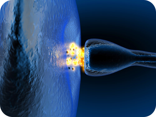
How does a nervous system signal move from one cell to the next?
It literally jumps by way of a chemical transmitter. Notice the two cells are not connected, but separated by a small gap. The synapse. The space between a neuron and the next cell.
Nerve Impulses
Nerve impulses are electrical in nature. They result from a difference in electrical charge across the plasma membrane of a neuron. How does this difference in electrical charge come about? The answer involves ions , which are electrically charged atoms or molecules.
Resting Potential
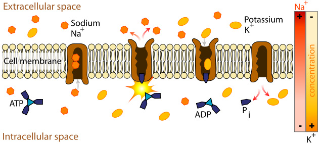
When a neuron is not actively transmitting a nerve impulse, it is in a resting state, ready to transmit a nerve impulse. During the resting state, the sodium-potassium pump maintains a difference in charge across the cell membrane (see Figure below). It uses energy in ATP to pump positive sodium ions (Na + ) out of the cell and potassium ions (K + ) into the cell. As a result, the inside of the neuron is negatively charged compared to the extracellular fluid surrounding the neuron. This is due to many more positively charged ions outside the cell compared to inside the cell. This difference in electrical charge is called the resting potential.
Action Potential
A nerve impulse is a sudden reversal of the electrical charge across the membrane of a resting neuron. The reversal of charge is called an action potential. It begins when the neuron receives a chemical signal from another cell. The signal causes gates in sodium ion channels to open, allowing positive sodium ions to flow back into the cell. As a result, the inside of the cell becomes positively charged compared to the outside of the cell. This reversal of charge ripples down the axon very rapidly as an electric current (see Figure below).
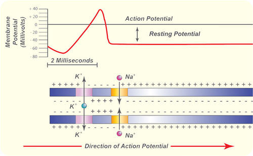
In neurons with myelin sheaths, ions flow across the membrane only at the nodes between sections of myelin. As a result, the action potential jumps along the axon membrane from node to node, rather than spreading smoothly along the entire membrane. This increases the speed at which it travels.
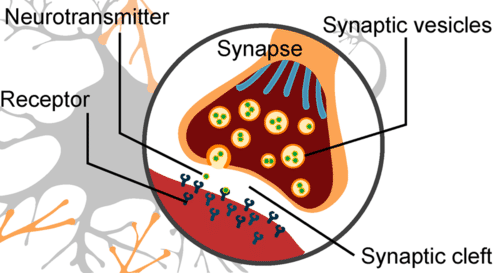
The place where an axon terminal meets another cell is called a synapse . The axon terminal and other cell are separated by a narrow space known as a synaptic cleft (see Figure below). When an action potential reaches the axon terminal, the axon terminal releases molecules of a chemical called a neurotransmitter . The neurotransmitter molecules travel across the synaptic cleft and bind to receptors on the membrane of the other cell. If the other cell is a neuron, this starts an action potential in the other cell.
- A nerve impulse begins when a neuron receives a chemical stimulus.
- The nerve impulse travels down the axon membrane as an electrical action potential to the axon terminal.
- The axon terminal releases neurotransmitters that carry the nerve impulse to the next cell.
- Define resting potential and action potential.
- Explain how resting potential is maintained
- Describe how an action potential occurs.
- What is a synapse?

- school Campus Bookshelves
- menu_book Bookshelves
- perm_media Learning Objects
- login Login
- how_to_reg Request Instructor Account
- hub Instructor Commons
- Download Page (PDF)
- Download Full Book (PDF)
- Periodic Table
- Physics Constants
- Scientific Calculator
- Reference & Cite
- Tools expand_more
- Readability
selected template will load here
This action is not available.

11.4: Neuronal Communication
- Last updated
- Save as PDF
- Page ID 22338

- Whitney Menefee, Julie Jenks, Chiara Mazzasette, & Kim-Leiloni Nguyen
- Reedley College, Butte College, Pasadena City College, & Mt. San Antonio College via ASCCC Open Educational Resources Initiative
By the end of this section, you will be able to:
- Explain the events that occur during the conduction of nerve impulses
- Differentiate the nerve impulse propagation in saltatory and continuous conduction
- Describe the components of synapses and compare electrical and chemical synapses
Having looked at the components of nervous tissue, and the basic anatomy of the nervous system, next comes an understanding of how nervous tissue is capable of communicating within the nervous system. Neurons communicate with other neurons, muscles or glands through the generation and conduction of nerve impulses. These nerve impulses represent changes in the electrical properties of the neuronal cell membrane. All cells have an electrical charge associated with their membrane. However, neurons and other cells are able to change their electrical charge by moving ions across the membrane. In this section, you will look at the basics of neuronal communication, mainly focusing on the conduction of the nerve impulse.
Conduction of Nerve Impulses
Neurons possess electrical excitability, which is the ability to respond to a stimulus by generating a nerve impulse. In the majority of cases, the dendrites of a neuron are the place where local changes in the membrane electrical properties happen through synapses. Dendrites receive stimuli from the external environment (e.g. somatic sensory neurons) or internal environment (e.g. visceral sensory neurons, motor neurons or interneurons). The amount of change in the membrane electrical charge is determined by the strength of the stimulus that causes it. For example, a needle pricking a fingertip will result in a bigger stimulus compared to a blunt object touching the same fingertip. Once a stimulus (or multiple stimuli) produces a significant change in the membrane electrical properties of a dendrite and reaches a predetermined threshold , then a nerve impulse (also called action potential) occurs. An action potential is generated at the axon hillock of a neuron and progresses rapidly along the axon's plasma membrane to reach the targets (a second neuron, a muscle or a gland). This movement of an action potential along the axon is called propagation . While the stimuli can be weak or strong, the action potential follows a All-or-None Law by which it always has the same strength (referred to as amplitude) independently of the stimulus. This minimizes the possibility that information will be lost along the way. The only way to modulate the response is through the frequency of action potentials - how many action potentials reach the target in a set amount of time. A bigger stimulus will produce a series (or train) of action potentials that are close together, while a weak stimulus will produce sparse action potentials.
The speed of an action potential is influenced by the diameter of the axon and by its myelination. The larger the diameter of the axon, the faster the action potential will be conducted. Myelinated axons are able to carry action potentials faster than unmyelinated axons. As discussed in the previous section, myelinating cells (oligodendrocytes in the CNS and Schwann cells in the PNS) wrap around axons forming the myelin. Nodes of Ranvier are gaps between segments of myelin. The electrical charges of the action potential can "jump" from one gap to another one, thus allowing a faster speed of the action potential. This progression of a nerve impulse is called saltatory conduction . However, in unmyelinated axons, one side of the axon is not covered by myelin and the electrical charges move along the entire axonal membrane, thus taking longer to reach their target. This progression of a nerve impulse is called continuous conduction . Once the action potential reaches the axon terminal, it is either transported as electrical charge into the next cell or transformed into a chemical signal, depending on the type of synapse that the synaptic end bulb is forming with its target.
Neurons and their targets form synapses. The neuron that generates and conducts the action potential to the target is called a presynaptic cell . The target cell receiving the action potential is called a postsynaptic cell . While the presynaptic cell is always a neuron (because only neurons have axons and can form a synapse), the postsynaptic cell can be a neuron or another type of cell such as skeletal, cardiac or smooth muscle cells, or glands. In Figure \(\PageIndex{1}\), a presynaptic neuron forms synapses with two postsynaptic neurons. The nerve impulse (or signal) travels from a presynaptic neuron to a postsynaptic cell. If the postsynaptic cell is a neuron, a new action potential might be generated in the postsynaptic neuron and reach its postsynaptic targets.

There are two types of connections between electrically active cells: electrical synapses and chemical synapses. In an electrical synapse , there is a direct connection between the presynaptic and postsynaptic cells and the connection is formed by gap junctions. Thus, the electrical charges of an action potential can pass directly from one cell to the next. If one cell delivers an action potential in an electrical synapse, the joined cell will also generate an action potential because the electrical charges will pass between the cells (Figure \(\PageIndex{2}\)). Although representing the minority of synapses, electrical synapses are found throughout the nervous system. These synapses also occur between excitable cells other than neurons, for example between smooth muscle cells in the intestines and cardiac muscle cells in the heart.

Chemical synapses involve the transmission of chemical information from one cell to the next and they represent the majority of the synapses found within the nervous system. In a chemical synapse , a chemical signal called a neurotransmitter , is released from the presynaptic cell and it affects the postsynaptic cell. There are many different types of neurotransmitters, for example acetylcholine, serotonin, dopamine, adrenaline, glutamate, etc. Each neurotransmitter has its own specific receptor on the postsynaptic membrane. Chemical synapses can then be classified based on the neurotransmitter that the cells use to communicate (for example glutamatergic synapses use glutamate). Different neurotransmitters and different receptors will determine the overall response to the stimulus. All chemical synapses have common characteristics, which can be summarized in this list and are shown in Figure \(\PageIndex{3}\):
- synaptic end bulb of presynaptic neuron
- neurotransmitter (packaged in vesicles)
- synaptic cleft
- receptors for neurotransmitter
- postsynaptic membrane of postsynaptic neuron
The synaptic end bulbs of chemical synapses are filled with vesicles containing one type of neurotransmitter. When an action potential reaches the axon terminals, the vesicles merge with the cell membrane at the synaptic end bulb, releasing the neurotransmitter through exocytosis into the small gap between the cells, known as the synaptic cleft . Once in the synaptic cleft, the neurotransmitter diffuses the short distance to the postsynaptic membrane and can interact with neurotransmitter receptors. Receptors are specific for the neurotransmitter, and the two fit together like a key and lock. One neurotransmitter binds to its receptor and will not bind to receptors for other neurotransmitters, making the binding a specific chemical event. The binding of the neurotransmitter to its receptor causes a brief electrical change across the postsynaptic membrane. The change depends on the type of neurotransmitter receptor. Changes in the postsynaptic cell membrane can cause a nerve impulse to begin in the postsynaptic cell or inhibit the generation of an action potential. The flow of information is unidirectional: from the presynaptic cell to the postsynaptic cell. After its release in a chemical synapse, neurotransmitters need to be removed from the synaptic cleft to ensure the propagation of new synaptic signals.

DISORDERS OF THE...
Nervous System: Alzheimer's and Parkinson's Disease
The underlying cause of some neurodegenerative diseases, such as Alzheimer’s and Parkinson’s, appears to be related to proteins—specifically, to proteins behaving badly. One of the strongest theories of what causes Alzheimer’s disease is based on the accumulation of beta-amyloid plaques, dense conglomerations of a protein that is not functioning correctly. Parkinson’s disease is linked to an increase in a protein known as alpha-synuclein that is toxic to the cells of the substantia nigra nucleus in the midbrain.
For proteins to function correctly, they are dependent on their three-dimensional shape. The linear sequence of amino acids folds into a three-dimensional shape that is based on the interactions between and among those amino acids. When the folding is disturbed, and proteins take on a different shape, they stop functioning correctly. But the disease is not necessarily the result of functional loss of these proteins; rather, these altered proteins start to accumulate and may become toxic. For example, in Alzheimer’s, the hallmark of the disease is the accumulation of these amyloid plaques in the cerebral cortex. The term coined to describe this sort of disease is “proteopathy” and it includes other diseases. Creutzfeld-Jacob disease, the human variant of the prion disease known as mad cow disease in the bovine, also involves the accumulation of amyloid plaques, similar to Alzheimer’s. Diseases of other organ systems can fall into this group as well, such as cystic fibrosis or type 2 diabetes. Recognizing the relationship between these diseases has suggested new therapeutic possibilities. Interfering with the accumulation of the proteins, and possibly as early as their original production within the cell, may unlock new ways to alleviate these devastating diseases.
CAREER CONNECTIONS
Neurophysiologist
Understanding how the nervous system works could be a driving force in your career. Studying neurophysiology is a very rewarding path to follow. It means that there is a lot of work to do, but the rewards are worth the effort.
The career path of a research scientist can be straightforward: college, graduate school, postdoctoral research, academic research position at a university. A Bachelor’s degree in science will get you started, and for neurophysiology that might be in biology, psychology, computer science, engineering, or neuroscience. But the real specialization comes in graduate school. There are many different programs out there to study the nervous system, not just neuroscience itself. Most graduate programs are doctoral, meaning that a Master’s degree is not part of the work. These are usually considered five-year programs, with the first two years dedicated to course work and finding a research mentor, and the last three years dedicated to finding a research topic and pursuing that with a near single-mindedness. The research will usually result in a few publications in scientific journals, which will make up the bulk of a doctoral dissertation. After graduating with a Ph.D., researchers will go on to find specialized work called a postdoctoral fellowship within established labs. In this position, a researcher starts to establish their own research career with the hopes of finding an academic position at a research university.
Other options are available if you are interested in how the nervous system works. Especially for neurophysiology, a medical degree might be more suitable so you can learn about the clinical applications of neurophysiology and possibly work with human subjects. An academic career is not a necessity. Biotechnology firms are eager to find motivated scientists ready to tackle the tough questions about how the nervous system works so that therapeutic chemicals can be tested on some of the most challenging disorders such as Alzheimer’s disease or Parkinson’s disease, or spinal cord injury.
Others with a medical degree and a specialization in neuroscience go on to work directly with patients, diagnosing and treating mental disorders. You can do this as a psychiatrist, a neuropsychologist, a neuroscience nurse, or a neurodiagnostic technician, among other possible career paths.
Concept Review
The basis of the electrical signal within a neuron is the action potential that propagates down the axon. For a neuron to generate an action potential, it needs to receive input from another source, either another neuron or a sensory stimulus. That input will result in a change in the electrical properties of the cell membrane, based on the strength of the stimulus. Once the stimulus is strong enough, it will generate an action potential that travels along the axon to the synaptic end bulb.
The diameter of the axon and the presence or absence of myelin determines how fast the action potential is conducted down the axon. A larger diameter and the presence of a myelin sheath will ensure a fast propagation of the action potential.
Synapses are the contacts between neurons, which can either be chemical or electrical in nature. Chemical synapses are far more common. At a chemical synapse, neurotransmitter is released from the presynaptic element and diffuses across the synaptic cleft. The neurotransmitter binds to a receptor protein and causes a change in the postsynaptic membrane (the PSP). The neurotransmitter must diffuse, be inactivated or removed from the synaptic cleft so that the stimulus is limited in time.
Review Questions
Q. At an electrical synapse, what do the presynaptic and postsynaptic cells communicate through?
A. neurotransmitters
B. neurotransmitter receptors
C. nodes of Ranvier
D. gap junctions
Q. Which of the following axons would propagate an action potential faster than the others?
A. myelinated, large diameter, axons
B. myelinated, small diameter, axons
C. unmyelinated, large diameter, axons
D. unmyelinated, small diameter, axons
Contributors and Attributions
OpenStax Anatomy & Physiology (CC BY 4.0). Access for free at https://openstax.org/books/anatomy-and-physiology
- Anatomy & Physiology
- Astrophysics
- Earth Science
- Environmental Science
- Organic Chemistry
- Precalculus
- Trigonometry
- English Grammar
- U.S. History
- World History
... and beyond
- Socratic Meta
- Featured Answers

Why can nerve impulses travel only in one direction?

When you get a nerve firing, you have probably heard that it is an electrical impulse that carries the signal. This is true, but it is not electrical in the same way your wall outlet works. This is electrochemical energy. Neurotransmitters are molecules that fit like a lock and key into a specific receptor. The receptor is located on the next cell in the line. When the neurotransmitter hits the receptor on the next cell in line, it signals that cell to begin a firing as well.
This will continue all the way down the length of the nerve track. In a nutshell, a nerve firing results in a chain reaction down the nerve cell's axon, or stemlike section. Sodium (Na+) ions flow in, potassium (K+) ions flow out, and we get an electrochemical gradient flowing down the length of the cell. You can think of it as a line of gunpowder that someone lit, with the flame traveling down the length of it. Common electrical power is more like a hose full of water, and when you put pressure on one end, the water shoots out the other.
Therefore, nerve impulses cannot travel in the opposite direction, because nerve cells only have neurotransmitter storage vesicles going one way, and receptors in one place.
https://answers.yahoo.com/question/index?qid=20080916232931AA4zPLL
Addendum to further questions: The impulses travel TO the CNS from the PNS. Do not confuse NERVE signals from the MUSCLE signals! Each goes ONE WAY, but together they coordinate to help use move. The following is a better description of the chemical processes involved: https://web.facebook.com/bryant.seabrooks/videos/10208641612752284/?t=325
Related questions
- Is aggression learned or innate?
- What is self-efficacy?
- What is the rationale for using adoption studies and twin studies in learning about genetic...
- Why are twin studies used to understand genetic contributions to human behavior?
- Describe how twin and adoption studies help us differentiate herediatary and enviormental on humans?
- Do adoption studies show that the personalities of adopted children match those of their...
- How does adoption and twin studies influence the nature vs. nurture debate?
- How do twin and adoption studies help us differentiate between the influences of nature and nurture?
- What is the differences between nicotinic and muscarinic receptor?
- What is acetylcholine classified as?
Impact of this question


Nerve Impulses: the Key to Understanding the Brain
Impulses are the basis of mind..
Posted October 17, 2019
One of the greatest, relatively underappreciated, discoveries in all of science was the discovery of the nerve impulse in the 1930s by the British Lord Adrian. Adrian did win a Nobel Prize for his discovery in 1932, but scholars underestimated its implications, which go beyond the fact that four later Nobel Prizes were awarded for work based on Adrian’s discovery. This included discovery of sodium and potassium ionic flux during impulses, the role of impulses in releasing neurotransmitters, and the role of membrane ion channels in impulse generation and second messenger cascades.
Like many discoveries in science, this one could not have been made without technological advances. Early studies with inferior technology had a very poor signal-to-noise ratio (not the thick baseline electrical noise in the illustration). Vast improvements in this ratio are obtained with today's technology and intracellular recording. In Adrian's day, the essential advance was the development of the capillary electrometer, which enabled the detection of very small electrical pulses on the order of one-millisecond duration. This instrumentation was crude and far inferior to later advances such as the oscilloscope and computer screens. Before Adrian’s use of the electrometer, scientists generally knew that peripheral nerves generated some kind of electrical signal, but nothing was known about the nature of the signal in individual neurons.
Nerves contain fibers from hundreds of neurons that produce a summed, relatively long duration and large wave that spreads down the nerve. No one knew how the individual nerve fibers contributed to this compound signal. Adrian answered this question by tedious microdissection of nerves into their individual fibers and recording stimulus-evoked responses in a single fiber. What Adrian saw was that the response was a series of voltage pulses, each about one millisecond long, all of the same amplitude in a given fiber. Decades later, the development of microelectrodes enabled confirmation of Adrian’s discovery in neurons in the brain.
This provided evidence of the basic similarity and difference between brains and the later development of computers. Both computers and brains convert the real world into representations. In computers, information is coded, in the form of 1s and 0s, and as nerve impulses in brains. Both computers and brains distribute and process this represented information, and can store it as memories. However, because brains are biological and use impulses to represent information, they can change their circuitry and can self-program. Unlike computers, brains also have will, including a likely degree of free will .
Brains have conspicuous functional states, ranging from intense conscious concentration to drowsiness, to sleep, to coma, to death. Neuronal electrical activity correlates in a systematic way with these state changes. The most conspicuous of these activity measures exist in terms of nerve impulse firing and the extracellular ionic currents they create at synapses, known as field potentials. As these field potentials reach the scalp, they produce the signal we call an electroencephalogram. Field potentials are technologically easier to record than individual nerve impulses, but more ambiguous to interpret because of the spatial summation of voltages from hundreds of heterogeneous neurons.
The original nerve impulse findings were that the rate of impulse firing governed the impact on neuronal targets, whether they be muscle or other neurons. Various labs, including my own, in the 1980s, discovered that the intervals between impulses also contained their own kind of information. For example, my lab reported that some neurons contained statistically significant serial ordering of impulse intervals in a neuron’s impulse stream. The intervals, at least in higher-level brain areas, are not random. They are serially dependent, as if they contained a message. If you are familiar with Markov transition probability, you can understand our finding that serial dependences exist in as many as five successive intervals (Sherry et al. 1982). This led us to suggest “byte processing” as a basic feature of neuronal information processing. This view has not caught on, and most people still seem to think that firing rate is the basic information code, despite the well-established temporal summation that occurs as impulses arrive at synapses. Bernard Katz demonstrated temporal summation of impulse effects in neuromuscular junctions in 1951 and later J.P. Segundo and colleagues confirmed it in neuronal synapses (Segundo et al., 1963).
It should not be surprising that there are serial dependencies in impulse intervals. For example, intracellular recording of postsynaptic potentials revealed that the polarization change caused by a single impulse input decays in a few milliseconds. However, a succession of closely spaced impulse inputs allows the polarization changes to summate.
These days, the emphasis needs to be put on impulse activity in defined circuitry. All neurons are linked in one or more circuits, and the impulse train in any one neuron is only a small part of the over-all circuit activity. The function of any given circuit depends on the circuit impulse pattern (CIP) of the whole circuit. Researchers have developed microelectrodes that allow recording of impulse trains from single neurons, but the problem is in implanting a series of electrodes so that each one monitors the activity of a selected neuron in a defined.
I think that research should focus on CIPs and the phase relationships of electrical activity among cortical circuits, both within and among cortical columns (Klemm, 2011). Nerve impulses have to be at the heart of consciousness, inasmuch as impulses contain the brain’s representation of information and create the synaptic field potentials.

We know from monitoring known anatomical pathways for specific sensations that the brain creates a CIP representation of the stimuli. As long as the CIPs remain active, the representation of sensation or neural processing is intact and may even be accessible to consciousness. However, if something disrupts ongoing CIPs to create a different set of CIPs, as for example would happen with a different stimulus, then the original representation disappears. If the original CIPs persist long enough, a memory could form, but otherwise, the information would be lost. The implication for memory formation is that the immediate period after learning must be protected from new inputs to keep the CIP representation of the learning intact long enough to form a more lasting memory.
Much current research shows that conscious awareness correlates with the degree of synchrony and time-locking of CIPs in various regions and within regions of cortex. The evidence comes from electroencephalographic monitoring of the oscillating field potentials in a given area. These are voltage waves that occur in multiple frequency bands. Phase relationships of voltage waves from different circuits surely reflect the timing of the impulse discharges that create those fields. I summarized the animal research evidence for this view in my first book, some 50 years ago (Klemm, 1969). Depending on the nature of the stimulus and mental state, these oscillations of various circuits may jitter with respect to each other or become time locked. The functional consequence of synchrony has to be substantial, and many others and I suggest that this is a fundamental aspect of consciousness. The correlation between frequency coherences and states of consciousness is clear. Frequency coherence reflects a “binding” of neurons into linked and shared electrochemical activity, but how this relates to conscious awareness will require a next great discovery in science.
Klemm, W. R. (1969). Animal Electroencephalography. New York: Academic Press.
Klemm, W. R. (2011). Atoms of Mind. The “Ghost in the Machine” Materializes. New York: Springer.
Segundo, J. P., et al. (1963). Sensitivity of neurons in Aplysia to temporal pattern of arriving impulses. J. Exp. Biol. 40: 643-667.
Sherry, C. J., Barrow, D. L., and Klemm, W. R. 1982. Serial dependencies and Markov processes of neuronal interspike intervals from rat cerebellum. Brain Res. Bull. 8: 163‑169.

William Klemm , Ph.D ., is a senior professor of Neuroscience at Texas A&M University.
- Find a Therapist
- Find a Treatment Center
- Find a Psychiatrist
- Find a Support Group
- Find Teletherapy
- United States
- Brooklyn, NY
- Chicago, IL
- Houston, TX
- Los Angeles, CA
- New York, NY
- Portland, OR
- San Diego, CA
- San Francisco, CA
- Seattle, WA
- Washington, DC
- Asperger's
- Bipolar Disorder
- Chronic Pain
- Eating Disorders
- Passive Aggression
- Personality
- Goal Setting
- Positive Psychology
- Stopping Smoking
- Low Sexual Desire
- Relationships
- Child Development
- Therapy Center NEW
- Diagnosis Dictionary
- Types of Therapy

Understanding what emotional intelligence looks like and the steps needed to improve it could light a path to a more emotionally adept world.
- Coronavirus Disease 2019
- Affective Forecasting
- Neuroscience

The “Coming Out” of the Electrical Synapse
- by an electrical synapse, in which the two cells touch and are connected by tiny holes, which lets the nerve impulse pass directly from one neuron to the other; or
- by a chemical synapse, where the two cells do not touch and the nerve impulse needs particular molecules to bridge the gap between them.
Chemical synapses are slower than electrical ones but are also far more flexible. This valuable flexibility is the foundation of all learning .

On either side of the synapse, the axon and dendrite have evolved specialized molecules to perform their respective tasks.

- school Campus Bookshelves
- menu_book Bookshelves
- perm_media Learning Objects
- login Login
- how_to_reg Request Instructor Account
- hub Instructor Commons
- Download Page (PDF)
- Download Full Book (PDF)
- Periodic Table
- Physics Constants
- Scientific Calculator
- Reference & Cite
- Tools expand_more
- Readability
selected template will load here
This action is not available.

35.4: How Neurons Communicate - Nerve Impulse Transmission within a Neuron- Resting Potential
- Last updated
- Save as PDF
- Page ID 13868

Learning Objectives
- Explain the formation of the resting potential in neurons
Nerve Impulse Transmission within a Neuron
For the nervous system to function, neurons must be able to send and receive signals. These signals are possible because each neuron has a charged cellular membrane (a voltage difference between the inside and the outside). The charge of this membrane can change in response to neurotransmitter molecules released from other neurons and environmental stimuli. Any voltage is a difference in electric potential between two points; for example, the separation of positive and negative electric charges on opposite sides of a resistive barrier. To understand how neurons communicate, one must first understand the basis of charged membranes and the baseline or ‘resting’ membrane charge.
Neuronal Charged Membranes
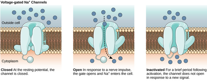
The lipid bilayer membrane that surrounds a neuron is impermeable to charged molecules or ions. To enter or exit the neuron, ions must pass through special proteins called ion channels that span the membrane. Ion channels have different configurations: open, closed, and inactive. Some ion channels need to be activated in order to open and allow ions to pass into or out of the cell. These ion channels are sensitive to the environment and can change their shape accordingly. Ion channels that change their structure in response to voltage changes are called voltage-gated ion channels. Voltage-gated ion channels regulate the relative concentrations of different ions inside and outside the cell. The difference in total charge between the inside and outside of the cell is called the membrane potential.
Resting Membrane Potential
For quiescent cells, the relatively-static membrane potential is known as the resting membrane potential. The resting membrane potential is at equilibrium since it relies on the constant expenditure of energy for its maintenance. It is dominated by the ionic species in the system that has the greatest conductance across the membrane. For most cells, this is potassium. As potassium is also the ion with the most-negative equilibrium potential, usually the resting potential can be no more negative than the potassium equilibrium potential.
A neuron at rest is negatively charged because the inside of a cell is approximately 70 millivolts more negative than the outside (−70 mV); this number varies by neuron type and by species. This voltage is called the resting membrane potential and is caused by differences in the concentrations of ions inside and outside the cell. If the membrane were equally permeable to all ions, each type of ion would flow across the membrane and the system would reach equilibrium. Because ions cannot simply cross the membrane at will, there are different concentrations of several ions inside and outside the cell. The difference in the number of positively-charged potassium ions (K + ) inside and outside the cell dominates the resting membrane potential. When the membrane is at rest, K + ions accumulate inside the cell due to a net movement with the concentration gradient. The negative resting membrane potential is created and maintained by increasing the concentration of cations outside the cell (in the extracellular fluid) relative to inside the cell (in the cytoplasm). The negative charge within the cell is created by the cell membrane being more permeable to K + movement than Na + movement.

In neurons, potassium ions (K+) are maintained at high concentrations within the cell, while sodium ions (Na+) are maintained at high concentrations outside of the cell. The cell possesses potassium and sodium leakage channels that allow the two cations to diffuse down their concentration gradient. However, the neurons have far more potassium leakage channels than sodium leakage channels. Therefore, potassium diffuses out of the cell at a much faster rate than sodium leaks in. More cations leaving the cell than entering it causes the interior of the cell to be negatively charged relative to the outside of the cell. The actions of the sodium-potassium pump help to maintain the resting potential, once it is established. Recall that sodium-potassium pumps bring two K + ions into the cell while removing three Na+ ions per ATP consumed. As more cations are expelled from the cell than are taken in, the inside of the cell remains negatively charged relative to the extracellular fluid.
- When the neuronal membrane is at rest, the resting potential is negative due to the accumulation of more sodium ions outside the cell than potassium ions inside the cell.
- Potassium ions diffuse out of the cell at a much faster rate than sodium ions diffuse into the cell because neurons have many more potassium leakage channels than sodium leakage channels.
- Sodium-potassium pumps move two potassium ions inside the cell as three sodium ions are pumped out to maintain the negatively-charged membrane inside the cell; this helps maintain the resting potential.
- ion channel : a protein complex or single protein that penetrates a cell membrane and catalyzes the passage of specific ions through that membrane
- membrane potential : the difference in electrical potential across the enclosing membrane of a cell
- resting potential : the nearly latent membrane potential of inactive cells


Turn Your Curiosity Into Discovery
Latest facts.
9 Facts About World Baking Day May 19th
8 Facts About International Tea Day May 21st
19 captivating facts about nerve impulse.
Written by Dayna Weintraub
Modified & Updated: 03 Mar 2024
Reviewed by Sherman Smith
- Membrane Potential Facts
- Neurons Facts
- Neurotransmitters Facts
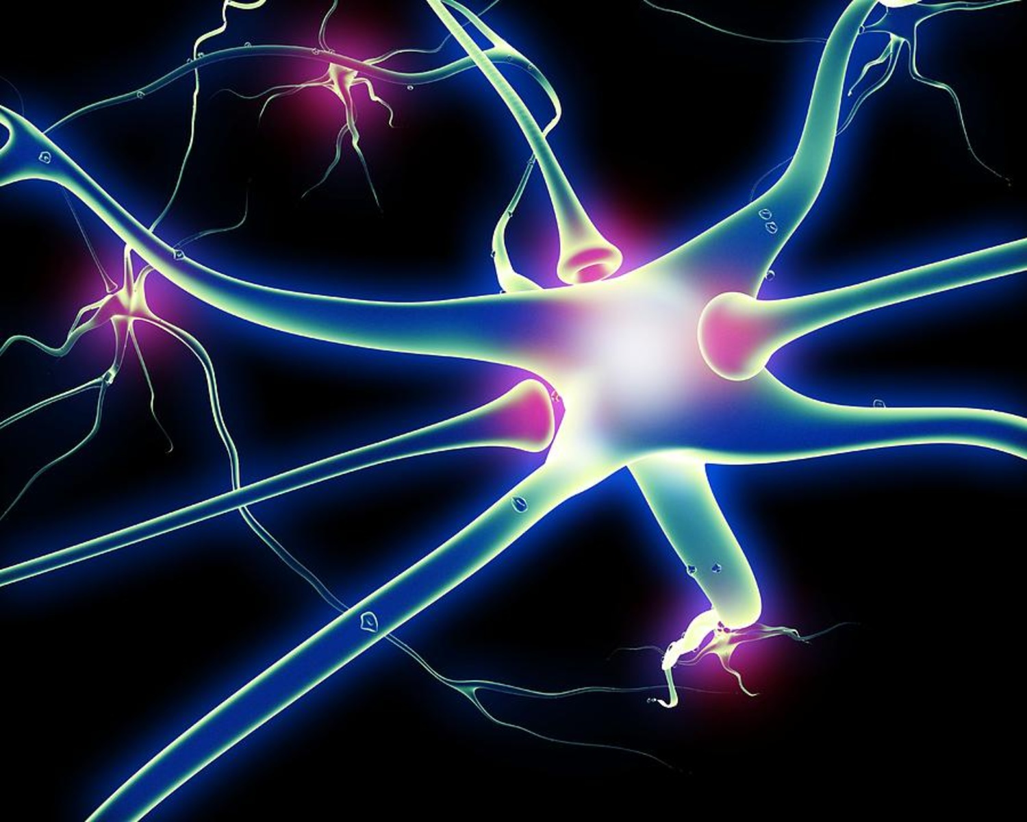
Nerve impulses are the electrical signals that allow information to travel throughout our bodies. They play a crucial role in our daily functioning, from enabling us to move our muscles to allowing us to feel sensations and perceive the world around us. Understanding how nerve impulses work is not only fascinating but also essential in the field of biology and neuroscience.
In this article, we will explore 19 captivating facts about nerve impulses that will deepen your understanding of this intricate biological process. From the minute details of how nerve cells communicate to the astonishing speed at which nerve impulses travel, we will delve into the world of neurons and the remarkable ways in which they function.
Get ready to embark on a journey through the fascinating realm of nerve impulses, as we unravel the mysteries behind these electrical signals and appreciate the complexity of the human nervous system.
Key Takeaways:
- Nerve impulses are like lightning bolts in our bodies, helping us move, feel, and react quickly. They’re super fast and essential for everything from muscle movements to experiencing the world through our senses.
- Understanding nerve impulses is crucial for developing treatments for conditions like multiple sclerosis and epilepsy. Scientists are unraveling their mysteries to improve human health and make groundbreaking medical advancements.
Nerve impulses are the result of electrical changes in neuron membranes.
When a neuron is stimulated, the electrical charge inside the neuron’s membrane changes, creating an electrical impulse that travels along the neuron.
Nerve impulses can travel at speeds of up to 120 meters per second.
Thanks to their incredible speed, nerve impulses allow us to react quickly to stimuli, such as pulling our hand away from a hot surface.
The myelin sheath increases the speed of nerve impulse transmission.
The myelin sheath, a fatty substance that wraps around some neurons, acts as an insulator and facilitates faster conduction of the electrical signal.
Nerve impulses can be either excitatory or inhibitory.
Excitatory nerve impulses stimulate the receiving neuron, while inhibitory impulses prevent the neuron from firing, balancing the overall neural activity.
The process of transmitting nerve impulses is called synaptic transmission.
At the synapse, the junction between two neurons, chemical messengers called neurotransmitters are released to transmit the signal from one neuron to another.
Nerve impulses can be both electrical and chemical in nature.
While the initial impulse within a neuron is electrical, the transmission of the impulse between neurons involves the release and reception of chemical neurotransmitters.
Sodium and potassium ions play a crucial role in nerve impulse transmission.
During the transmission, there is an exchange of sodium and potassium ions across the neuron’s membrane, creating the necessary electrical changes for the impulse to travel.
Nerve impulses can travel in both directions within a neuron.
While the majority of nerve impulses travel in one direction, certain types of neurons allow for bidirectional transmission, enabling more complex neural pathways.
Reflex actions are a result of fast, involuntary nerve impulse transmission.
Reflexes, such as pulling our hand away from a sharp object, occur due to a rapid nerve impulse bypassing the brain and directly triggering a motor response.
Nerve impulses can be amplified and modulated in the brain.
The brain has the remarkable ability to regulate and modify nerve impulses, allowing for intricate processing and interpretation of sensory information.
Nerve impulses are essential for muscle movement and coordination.
From the simplest movements to complex athletic performances, nerve impulses are responsible for initiating and controlling muscle contractions.
The speed of nerve impulse transmission can be affected by various factors.
Temperature, myelin thickness, and the diameter of the nerve fiber can all impact the speed at which a nerve impulse travels.
Chemical substances can alter or block nerve impulse transmission.
Anesthetics and certain drugs can temporarily disrupt nerve impulse transmission by interfering with ion channels or neurotransmitter release .
Nerve impulses play a crucial role in sensory perception.
By transmitting signals from receptors in our sensory organs to the brain, nerve impulses enable us to experience the world through our senses.
Abnormal nerve impulse transmission can lead to neurological disorders.
Conditions such as multiple sclerosis, Parkinson’s disease, and epilepsy are all characterized by disrupted or faulty nerve impulse transmission.
Nerve impulses can be studied through electrophysiology.
Electrophysiology is a branch of science that focuses on the electrical properties and activity of living cells, including the measurement of nerve impulses.
Nerve impulse transmission requires energy.
The active transport of ions and the maintenance of concentration gradients during nerve impulse transmission require a significant amount of cellular energy.
The human brain generates millions of nerve impulses every second.
With billions of neurons firing in the brain at any given moment, the sheer magnitude of nerve impulses being transmitted is awe-inspiring.
Understanding nerve impulses is crucial for advancing medical treatments.
By unraveling the mysteries of nerve impulses, scientists and researchers can develop innovative therapies for neurological disorders and improve overall human health.
In Conclusion
The 19 captivating facts about nerve impulses highlight the remarkable complexity and importance of these electrical signals in our bodies. From the rapid transmission of reflex actions to the intricate processing of sensory information in the brain, nerve impulses play a vital role in our everyday functioning. By delving into the fascinating world of nerve impulses, we gain a deeper appreciation for the incredible workings of the human nervous system .
In conclusion, the process of nerve impulse transmission is truly fascinating and essential for the functioning of our body. From the intricate network of neurons to the complex electrical signals, every aspect plays a crucial role in ensuring the transmission of information throughout our nervous system.We have learned that nerve impulses travel at incredible speeds, allowing for quick responses to external stimuli. The myelin sheath, with its insulating properties, facilitates the rapid conduction of impulses along the axons. Additionally, the actions of ion channels and neurotransmitters ensure the precise and efficient transmission of signals between neurons.Understanding the intricate mechanisms behind nerve impulse transmission not only enhances our knowledge of the human body but also helps in the development of treatments for neurological disorders. The more we delve into the complexities of nerve impulses, the better equipped we become to unlock the mysteries of the nervous system.
1. What is a nerve impulse?
A nerve impulse is an electrical signal that is transmitted through neurons, allowing for communication within the nervous system.
2. How fast does a nerve impulse travel?
A nerve impulse can travel at speeds of up to 120 meters per second, depending on factors such as the diameter of the nerve fiber and the presence of a myelin sheath.
3. What is the role of the myelin sheath in nerve impulse transmission?
The myelin sheath acts as an insulating layer around the axons of some neurons, enabling faster transmission of nerve impulses and preventing the signal from dissipating.
4. How do nerve impulses cross the synapse?
Nerve impulses are transmitted across the synapse through the release of neurotransmitters. These chemical messengers travel from the axon of one neuron to the dendrite of the next, facilitating transmission of the signal.
5. What happens when there is a disruption in nerve impulse transmission?
Disruptions in nerve impulse transmission can lead to various neurological disorders, such as multiple sclerosis or peripheral neuropathy, which can affect motor coordination, sensation, and other bodily functions.
6. Can nerve impulses be consciously controlled?
While involuntary nerve impulses regulate essential body functions, some nerve impulses can be consciously controlled, allowing us to move our muscles voluntarily and perform complex actions.
7. Are all nerve impulses the same?
No, nerve impulses can vary in strength and frequency, depending on the intensity and nature of the stimulus received by the sensory receptors.
8. How does the brain interpret nerve impulses?
The brain interprets nerve impulses based on their strength, frequency, and the specific neural pathways they follow. This interpretation allows us to perceive and react to different sensory stimuli.
9. Can nerve impulses travel in both directions?
While most nerve impulses travel in one direction, from the dendrites to the axon terminals, certain nerves, such as those involved in reflexes, can transmit impulses in both directions.
10. Can nerve impulses be artificially stimulated?
Yes, with the help of various technologies and medical interventions, such as electrical stimulation and nerve implants, it is possible to artificially stimulate nerve impulses in certain situations for therapeutic purposes.
Was this page helpful?
Our commitment to delivering trustworthy and engaging content is at the heart of what we do. Each fact on our site is contributed by real users like you, bringing a wealth of diverse insights and information. To ensure the highest standards of accuracy and reliability, our dedicated editors meticulously review each submission. This process guarantees that the facts we share are not only fascinating but also credible. Trust in our commitment to quality and authenticity as you explore and learn with us.
Share this Fact:
Why can nerve impulses travel only in one direction?
Nerve impulse: neurons communicate with one another through nerve impulses. nerve impulses are electrical signals that travel along dendrites onward the segment of an axon filament. the movement of ions into and out of the cell produces the action potential. cause of impulse transmission in one direction: nerve impulses only travel in one direction in neurons. this impulse transmission is dependent on synaptic transmission. this happens because nerve cells only have one transmission site. the receptors also work in one direction. the nerve impulse works on the principle of depolarization and repolarization. the nerve cells only have neurotransmitter storage vesicles in one way. nerve impulses must cross synaptic junctions to travel from one cell to another. the nerve cells are lined up in a long track, with the head of one cell connecting to the tail of another. there are tiny gaps between these cells. these tiny gaps are referred to as nerve synapses. when a nerve fires, an action potential is generated along the nerve track. the sodium-potassium pump ejects three sodium ions for every two potassium ions pumped into the cell; energy is needed for this process. it results in an action potential. because neurotransmitter storage vesicles and receptors are present in one location, impulse transmission occurs only in one direction..

At what speed do nerve impulses travel in mammals?


IMAGES
VIDEO
COMMENTS
100. Figure 42.2.2 42.2. 2: The (a) resting membrane potential is a result of different concentrations of Na + and K + ions inside and outside the cell. A nerve impulse causes Na + to enter the cell, resulting in (b) depolarization. At the peak action potential, K + channels open and the cell becomes (c) hyperpolarized.
For an action potential to communicate information to another neuron, it must travel along the axon and reach the axon terminals where it can initiate neurotransmitter release. The speed of conduction of an action potential along an axon is influenced by both the diameter of the axon and the axon's resistance to current leak.
When the buildup of charge was great enough, a sudden discharge of electricity occurred. A nerve impulse is similar to a lightning strike. Both a nerve impulse and a lightning strike occur because of differences in electrical charge, and both result in an electric current. Figure 11.4.1 11.4. 1: Lightning.
Neurotransmitters are how we communicate between one cell and the next. Synapses between neurons are either excitatory or inhibitory - and that all comes down to the neurotransmitter released. Excitatory neurotransmitters cause the signal to propagate - more action potentials are triggered. Inhibitory signals work to cancel the signal.
It can fire nerve impulses, or action potentials. And it can carry out the metabolic processes required to stay alive. ... In many cases, they can carry current in both directions so that depolarization of a postsynaptic neuron will lead to depolarization of a presynaptic neuron. This kind of bends the definitions of presynaptic and ...
A single neuron may have more than one set of dendrites, and may receive many thousands of input signals. Whether or not a neuron is excited into firing an impulse depends on the sum of all of the excitatory and inhibitory signals it receives. If the neuron does end up firing, the nerve impulse, or action potential, is conducted down the axon.
Nerve impulses travel just as fast through the network of nerves inside the body. Figure 11.41.1 11.41. 1: The axons of many neurons, like the one shown here, are covered with a fatty layer called myelin sheath. The sheath covers the axon, like the plastic covering on an electrical wire, and allows nerve impulses to travel faster along the axon.
A nerve impulse is a sudden reversal of the electrical charge across the membrane of a resting neuron. The reversal of charge is called an action potential. It begins when the neuron receives a chemical signal from another cell. The signal causes gates in sodium ion channels to open, allowing positive sodium ions to flow back into the cell.
11.4: Neuronal Communication. Having looked at the components of nervous tissue, and the basic anatomy of the nervous system, next comes an understanding of how nervous tissue is capable of communicating within the nervous system. Neurons communicate with other neurons, muscles or glands through the generation and conduction of nerve impulses.
The best generic answer from a 2008 post follows (with my own edits for clarity): A Nerve electrical impulse only travels in one direction. There are several reasons nerve impulses only travel in one direction. The most important is synaptic transport. In order for a "nerve impulse" to pass from cell to cell, it must cross synaptic junctions.
In computers, information is coded, in the form of 1s and 0s, and as nerve impulses in brains. Both computers and brains distribute and process this represented information, and can store it as ...
In a chemical synapse, a nerve impulse can travel in only one direction. In contrast, in an electrical synapse, the impulse travels in both directions. Also, across a chemical synapse, the impulse is transmitted with a 0.5-millisecond delay, while across an electrical synapse, the delay is almost non-existent.
Action Potential. A nerve impulse is a sudden reversal of the electrical charge across the membrane of a resting neuron. The reversal of charge is called an action potential. It begins when the neuron receives a chemical signal from another cell. The signal causes gates in sodium ion channels to open, allowing positive sodium ions to flow back into the cell.
Saltatory conduction. In neuroscience, nerve conduction velocity (CV) is the speed at which an electrochemical impulse propagates down a neural pathway.Conduction velocities are affected by a wide array of factors, which include age, sex, and various medical conditions. Studies allow for better diagnoses of various neuropathies, especially demyelinating diseases as these conditions result in ...
A nerve impulse is initiated by a stimulus, that is, a change in the internal or external environment. This stimulus triggers a receptor to send a nerve impulse to our central nervous system (CNS). The CNS, consisting of the brain and spinal cord, processes the information. Nerve impulses are then transmitted from the CNS to different organs ...
Nerve impulses travel from one neuron to another through a process called synaptic transmission. The impulse reaches the end of one neuron, the presynaptic neuron, and triggers the release of neurotransmitters. These chemicals cross the synaptic gap and bind to receptors on the next neuron, the postsynaptic neuron. This binding generates a new impulse in the postsynaptic neuron, continuing the ...
A nerve cell can receive both EPSPs and IPSPs at the same time. The likelihood of the cell firing is determined by adding up the excitatory and the inhibitory synaptic input. ... There are several reasons nerve impulses only travel in one direction. The most important is synaptic transport. For a "nerve impulse" to pass from cell to cell, it ...
If you cut a piece of that axon, rotate it 180 degrees and join it back in the cutting points, it will conduct the action potential the same way. That being said, imagine that you stimulate that axon at a given point. There will be two action potentials, going to opposite ways: unmyelinated (A) and myelinated (B) nerve cells.
A nerve impulse causes Na+ to enter the cell, resulting in (b) depolarization. At the peak action potential, K+ channels open and the cell becomes (c) hyperpolarized. In neurons, potassium ions (K+) are maintained at high concentrations within the cell, while sodium ions (Na+) are maintained at high concentrations outside of the cell. The cell ...
Nerve impulses can travel in both directions within a neuron. While the majority of nerve impulses travel in one direction, certain types of neurons allow for bidirectional transmission, enabling more complex neural pathways. Reflex actions are a result of fast, involuntary nerve impulse transmission.
Neurons communicate with one another through nerve impulses. Nerve impulses are electrical signals that travel along dendrites onward the segment of an axon filament. The movement of ions into and out of the cell produces the action potential. Cause of impulse transmission in one direction: Nerve impulses only travel in one direction in neurons.
A. False In axons, impulses travel in both directions from a point of electrical stimulation.. B. True The transmitter vesicles are in the pre-synaptic terminal.. C. False Impulse propagation is a different process and much slower than electrical current.. D. True Nerves cannot be re-excited while their membrane polarity is reversed.. E. False The shorter refractory periods in somatic nerves ...